The sympathetic nervous system is a critical component of the autonomic nervous system, orchestrating the body’s rapid response to stress through a complex network of neural pathways. This diagram illustrates the diverse ways preganglionic neurons from the spinal cord connect to ganglia and target effectors, highlighting the versatility of the sympathetic division in maintaining physiological balance.
Labels in the Diagram
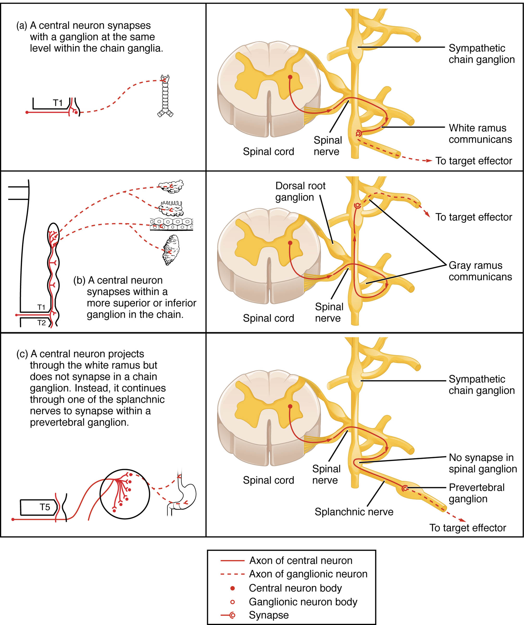
T1 The T1 label marks the first thoracic segment of the spinal cord, where preganglionic neurons originate. These neurons project axons to initiate sympathetic responses at this level or beyond.
T2 The T2 segment, the second thoracic level, serves as another origin point for preganglionic fibers. It demonstrates how neurons can extend to superior or inferior ganglia within the chain.
T5 The T5 segment, located in the mid-thoracic region, shows a neuron projecting through a splanchnic nerve. This pathway leads to prevertebral ganglia, bypassing the chain ganglia.
Spinal Cord The Spinal Cord is the central structure housing preganglionic neuron cell bodies in the lateral horn. It serves as the starting point for sympathetic signals that travel to peripheral ganglia.
Spinal Nerve The Spinal Nerve emerges from the spinal cord, carrying both sensory and motor fibers. It includes the white and gray rami communicantes that connect to the sympathetic chain ganglion.
Dorsal Root Ganglion The Dorsal Root Ganglion contains sensory neuron cell bodies, located near the spinal nerve. It does not directly participate in sympathetic function but is part of the overall spinal nerve complex.
Sympathetic Chain Ganglion The Sympathetic Chain Ganglion is a paravertebral ganglion where preganglionic neurons synapse with postganglionic neurons. It facilitates the relay of sympathetic signals to target effectors.
White Ramus Communicans The White Ramus Communicans is a preganglionic fiber bundle covered with myelin, connecting the spinal nerve to the sympathetic chain ganglion. It carries axons from the spinal cord to the ganglia.
Gray Ramus Communicans The Gray Ramus Communicans consists of unmyelinated postganglionic fibers returning from the sympathetic chain ganglion to the spinal nerve. It distributes signals to nearby effectors like blood vessels and sweat glands.
Splanchnic Nerve The Splanchnic Nerve carries preganglionic fibers that bypass the sympathetic chain ganglion to reach prevertebral ganglia. It innervates abdominal organs such as the stomach and intestines.
Prevertebral Ganglion The Prevertebral Ganglion is a collateral ganglion located anterior to the vertebral column, such as the celiac or superior mesenteric ganglion. It receives splanchnic nerve input and synapses with postganglionic neurons.
To Target Effector The To Target Effector label indicates the final destination of postganglionic fibers, which innervate organs like the heart, lungs, or sweat glands. This completes the sympathetic pathway, triggering physiological responses.
Axon of Central Neuron The Axon of Central Neuron represents the preganglionic fiber originating from the spinal cord. It varies in its synaptic destination depending on the pathway taken.
Axon of Ganglionic Neuron The Axon of Ganglionic Neuron is the postganglionic fiber that extends from the ganglion to the target effector. It releases norepinephrine to activate the effector organ.
Central Neuron Body The Central Neuron Body is the cell body of the preganglionic neuron located in the lateral horn of the spinal cord. It initiates the sympathetic response by sending axons outward.
Ganglionic Neuron Body The Ganglionic Neuron Body resides within the sympathetic chain ganglion or prevertebral ganglion. It receives the preganglionic signal and relays it to the effector.
Synapse The Synapse is the junction where the preganglionic neuron communicates with the ganglionic neuron. This connection uses acetylcholine to transmit the signal.
Anatomy of Sympathetic Neural Pathways
The sympathetic division begins in the thoracic and lumbar regions of the spinal cord, where preganglionic neurons extend axons through the white ramus communicans. These fibers may synapse within the sympathetic chain ganglion at the same level, travel to a superior or inferior ganglion, or project via the splanchnic nerve to a prevertebral ganglion.
- Lateral Horn: Contains central neuron bodies that initiate sympathetic signals.
- Paravertebral Ganglia: Form the sympathetic chain ganglion along the spinal column.
- Prevertebral Ganglia: Include structures like the celiac ganglion for abdominal control.
This flexibility allows the system to efficiently distribute signals to diverse effectors throughout the body.
Physiological Roles of Sympathetic Connections
The sympathetic chain ganglion serves as a hub for coordinating the body’s fight-or-flight response. Preganglionic fibers from the spinal nerve synapse here, with postganglionic fibers traveling via the gray ramus communicans to effectors.
- Heart Rate Increase: Stimulates the heart to pump faster during stress.
- Sweat Gland Activation: Regulates temperature through skin innervation.
- Blood Vessel Constriction: Redirects blood flow to essential muscles.
The splanchnic nerve pathway, leading to the prevertebral ganglion, controls abdominal organs, ensuring energy mobilization during emergencies.
Variations in Synaptic Pathways
The dorsal root ganglion provides sensory input but does not synapse in the sympathetic pathway. In contrast, the T1, T2, and T5 levels illustrate different synaptic options, showcasing the system’s adaptability.
- Same-Level Synapse: Occurs when the axon of central neuron meets a ganglionic neuron body at the same spinal level.
- Vertical Synapse: Allows projection to a higher or lower sympathetic chain ganglion.
- Splanchnic Route: Bypasses the chain to reach a prevertebral ganglion for visceral control.
This variability ensures comprehensive coverage of the body’s needs during sympathetic activation.
Clinical Significance of Sympathetic Pathways
Understanding these connections aids in diagnosing autonomic disorders related to the sympathetic division. Damage to the spinal cord or splanchnic nerve can disrupt normal function, leading to issues like irregular heart rate or poor digestion.
- Horner’s Syndrome: Results from interrupted superior cervical ganglion pathways, causing ptosis and miosis.
- Autonomic Dysreflexia: Overactivity due to spinal cord injury above T6.
- Therapeutic Interventions: Nerve blocks target prevertebral ganglia for pain relief.
Accurate mapping of these pathways supports effective treatment strategies.
Evolutionary and Research Perspectives
The sympathetic division evolved to enhance survival by rapidly mobilizing resources via the spinal nerve and sympathetic chain ganglion. Ongoing research explores how these pathways adapt to modern stressors, informing medical advancements.
- Evolutionary Advantage: Quick responses via the splanchnic nerve supported ancient survival needs.
- Neuroimaging Studies: Reveal synapse efficiency in the prevertebral ganglion.
- Future Applications: Could improve treatments for autonomic dysfunction.
This knowledge continues to deepen our understanding of human physiology.
In conclusion, the sympathetic division demonstrates a sophisticated network of neural connections, from the spinal cord to target effectors via the sympathetic chain ganglion and prevertebral ganglion. This diagram provides a clear foundation for exploring its anatomy and physiological significance, offering valuable insights for clinical practice and research.

