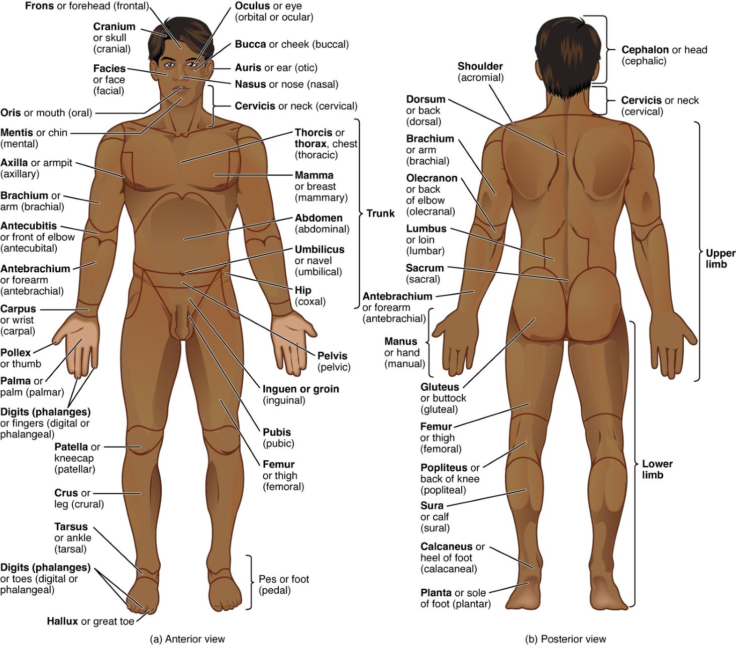
The human body is a complex structure, meticulously divided into various regions that are essential for anatomical study and medical practice. This image presents the Frons or Forehead, Cranium or Skull, Facies or Face, Oris or Mouth, Mentis or Chin, Auricula or Ear, Nasus or Nose, Cervicis or Neck, Thoracis or Thorax, Mamma or Breast, Abdomen, Umbilicus or Navel, Hip, Pelvis, Inguen or Groin, Pubis, Femur or Thigh, Patella or Kneecap, Crus or Leg, Tarsus or Ankle, Pes or Foot, Hallux or Great Toe, Digiti or Fingers, Palma or Palm, Pollex or Thumb, Axilla or Armpit, Brachium or Arm, Antebrachium or Forearm, Antecubital or Front of Elbow, Carpus or Wrist, Dorsum or Back, Shoulder, Olecranon or Back of Elbow, Lumbus or Loin, Sacrum, Gluteus or Buttock, Popliteus or Back of Knee, Sura or Calf, Calcaneus or Heel, Planta or Sole of Foot, Cephalon or Head, Upper Limb, Lower Limb, and Manus or Hand in both anterior and posterior perspectives. Understanding these regions enhances the ability to locate and assess the body’s structures with precision.
Label Introductions:
- Frons or Forehead: The frons or forehead forms the upper part of the face, protecting the frontal lobe of the brain. It also contains sweat glands that help regulate body temperature.
- Cranium or Skull: The cranium or skull encases the brain, providing a hard protective shell. It consists of several bones fused together to support facial structures.
- Facies or Face: The facies or face includes features like the eyes, nose, and mouth, serving as the primary site for sensory input. It plays a key role in communication and expression.
- Oris or Mouth: The oris or mouth is the entry point for food and air, housing teeth and the tongue for digestion and speech. It is surrounded by muscles that facilitate chewing and articulation.
- Mentis or Chin: The mentis or chin provides structural support to the lower face, anchoring facial muscles. It also contributes to the overall contour of the jawline.
- Auricula or Ear: The auricula or ear collects sound waves, directing them into the ear canal for hearing. It also helps in maintaining balance through its inner structures.
- Nasus or Nose: The nasus or nose filters and warms inhaled air, aiding respiration. It contains olfactory receptors for the sense of smell.
- Cervicis or Neck: The cervicis or neck supports the head and houses major blood vessels and the trachea. It allows for a wide range of head movements.
- Thoracis or Thorax: The thoracis or thorax protects the heart and lungs within the rib cage. It expands and contracts to facilitate breathing.
- Mamma or Breast: The mamma or breast contains mammary glands for lactation in females. It also provides a layer of fat for cushioning.
- Abdomen: The abdomen contains digestive organs like the stomach and liver, protected by abdominal muscles. It plays a central role in metabolism and nutrient processing.
- Umbilicus or Navel: The umbilicus or navel marks the site of fetal attachment, serving as a scar. It has no significant physiological function in adults.
- Hip: The hip connects the lower limb to the pelvis, supporting body weight during movement. It allows for a wide range of motion and stability.
- Pelvis: The pelvis supports the lower abdominal organs and facilitates childbirth in females. It forms a bony ring that connects the spine to the legs.
- Inguen or Groin: The inguen or groin is the junction between the abdomen and thigh, housing major blood vessels and nerves. It is a common site for hernias.
- Pubis: The pubis forms the anterior part of the pelvis, supporting the bladder and reproductive organs. It also contributes to the pelvic girdle’s stability.
- Femur or Thigh: The femur or thigh is the longest bone, supporting body weight and enabling locomotion. It connects the knee to the hip joint.
- Patella or Kneecap: The patella or kneecap protects the knee joint, improving leverage for leg extension. It is a sesamoid bone embedded in the quadriceps tendon.
- Crus or Leg: The crus or leg extends from the knee to the ankle, housing muscles for movement. It supports the body’s weight during standing and walking.
- Tarsus or Ankle: The tarsus or ankle connects the foot to the leg, providing flexibility and stability. It consists of seven tarsal bones for weight distribution.
- Pes or Foot: The pes or foot supports the body’s weight and facilitates movement. It contains 26 bones, including the arches for shock absorption.
- Hallux or Great Toe: The hallux or great toe enhances balance and propulsion during walking. It has two phalanges, unlike the other toes.
- Digiti or Fingers: The digiti or fingers enable precise movements and grasping, supported by multiple phalanges. They contain flexor and extensor muscles for dexterity.
- Palma or Palm: The palma or palm forms the inner surface of the hand, aiding in gripping and sensation. It contains flexor tendons and sensory nerves.
- Pollex or Thumb: The pollex or thumb allows for opposition, essential for tool use and fine motor skills. It has a unique saddle joint for flexibility.
- Axilla or Armpit: The axilla or armpit contains lymph nodes and major blood vessels, aiding immune function. It allows arm movement and flexibility.
- Brachium or Arm: The brachium or arm extends from the shoulder to the elbow, housing the humerus bone. It enables lifting and reaching motions.
- Antebrachium or Forearm: The antebrachium or forearm contains the radius and ulna, supporting wrist and hand movements. It rotates to allow supination and pronation.
- Antecubital or Front of Elbow: The antecubital or front of elbow is a common site for venipuncture due to accessible veins. It connects the arm to the forearm.
- Carpus or Wrist: The carpus or wrist links the hand to the forearm, providing flexibility. It comprises eight carpal bones for movement and strength.
- Dorsum or Back: The dorsum or back supports the spinal column and houses back muscles. It protects the spinal cord and maintains posture.
- Shoulder: The shoulder connects the upper limb to the torso, allowing a wide range of motion. It comprises the clavicle and scapula for stability.
- Olecranon or Back of Elbow: The olecranon or back of elbow forms the bony prominence of the ulna. It stabilizes the elbow during extension.
- Lumbus or Loin: The lumbus or loin is the lower back region, supporting the spine. It contains muscles for posture and movement.
- Sacrum: The sacrum forms the base of the spine, connecting to the pelvis. It transmits weight from the upper body to the legs.
- Gluteus or Buttock: The gluteus or buttock contains large muscles for hip extension and stability. It cushions the pelvis during sitting.
- Popliteus or Back of Knee: The popliteus or back of knee contains tendons and ligaments for knee stability. It aids in knee flexion and rotation.
- Sura or Calf: The sura or calf houses muscles for ankle movement and propulsion. It supports walking and running activities.
- Calcaneus or Heel: The calcaneus or heel is the largest tarsal bone, absorbing impact during walking. It anchors the Achilles tendon for movement.
- Planta or Sole of Foot: The planta or sole of foot provides a stable base for standing. It contains fat pads for shock absorption.
- Cephalon or Head: The cephalon or head houses the brain and sensory organs, protected by the skull. It is the control center for bodily functions.
- Upper Limb: The upper limb includes the arm, forearm, and hand, enabling complex movements. It is supported by the shoulder girdle.
- Lower Limb: The lower limb comprises the thigh, leg, and foot, supporting body weight. It facilitates locomotion and balance.
- Manus or Hand: The manus or hand allows for grasping and manipulation, containing 27 bones. It is essential for daily activities and dexterity.
Introduction to Body Regions
The human body is divided into specific regions, each with unique anatomical features and functions. These regions, illustrated in anterior and posterior views, provide a roadmap for locating structures and understanding their roles. Familiarity with these areas is crucial for accurate diagnosis and treatment.
- Serve as reference points for medical examinations and imaging.
- Enable precise communication among healthcare professionals.
- Reflect the body’s symmetry and functional organization.
- Support the study of surface anatomy for clinical practice.
Anterior View: Frontal Body Regions
The anterior view highlights the front of the body, showcasing key regions like the thoracis or thorax and abdomen. This perspective is vital for assessing the chest, where the heart and lungs reside, and the abdominal area, home to digestive organs. It offers a clear view for examining superficial and deep structures.
- The thoracis region expands during breathing, driven by the diaphragm.
- The abdomen houses the liver, which metabolizes nutrients and detoxifies blood.
- The inguen area is a critical junction for vascular and nerve pathways.
- The pubis supports the bladder, influencing urinary function.
Posterior View: Back Body Regions
The posterior view focuses on the back, emphasizing regions like the dorsum or back and lumbus or loin. This view is essential for studying the spine, which protects the spinal cord, and the gluteus, which powers hip movement. It aids in evaluating posture and musculoskeletal health.
- The dorsum supports the vertebral column for spinal alignment.
- The lumbus contains the erector spinae muscles for posture.
- The sacrum connects the spine to the pelvis, distributing weight.
- The sura muscles propel the body during walking or running.
Upper and Lower Limbs: Functional Divisions
The upper limb and lower limb are specialized for mobility and support. The upper limb, including the brachium and manus, enables intricate tasks like writing, while the lower limb, with the femur and pes, bears weight and facilitates locomotion. These regions are critical for physical activity and stability.
- The brachium contains the humerus, supporting arm strength.
- The femur is the strongest bone, crucial for weight-bearing.
- The carpus allows wrist flexibility for hand movements.
- The tarsus stabilizes the foot for balance and propulsion.
Head and Neck: Sensory and Structural Hub
The cephalon or head and cervicis or neck are central to sensory and structural functions. The cranium protects the brain, while the cervicis houses the carotid arteries for blood supply. These areas are key for neurological and vascular assessments.
- The cranium encases the brain, regulating cognitive functions.
- The cervicis supports head rotation and houses the larynx.
- The facies contains the eyes, aiding visual perception.
- The auricula directs sound to the inner ear for hearing.
Conclusion
Exploring the regions of the human body through anterior and posterior views reveals the intricate design of its anatomy. The thoracis or thorax, abdomen, dorsum or back, and lower limb each contribute to the body’s functionality and resilience. This knowledge empowers professionals to navigate the body’s landscape, enhancing diagnostic precision and therapeutic approaches.

