The human body offers several key pulse sites where the heartbeat can be palpated, providing valuable insights into cardiovascular health. This diagram highlights the primary arterial locations where pulse can be measured, with the radial artery being the most commonly used due to its accessibility and reliability.
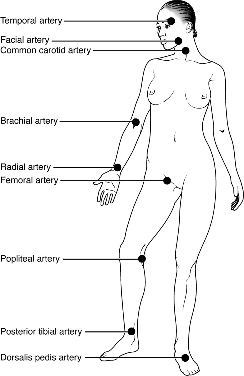
Temporal artery Temporal artery runs along the side of the head near the temple, making it a useful site for assessing pulse in specific clinical scenarios. Its superficial location allows for easy palpation, though it requires gentle pressure to avoid discomfort.
Facial artery Facial artery is located along the jawline and can be felt near the corner of the mouth. This site is less commonly used but can be an alternative when other pulse points are inaccessible.
Common carotid artery Common carotid artery lies on either side of the neck and is a major vessel carrying blood to the brain. Palpating this artery provides a strong pulse, often used in emergency situations to check circulation.
Brachial artery Brachial artery is found in the upper arm, commonly used to measure blood pressure with a cuff. Its pulse is palpable just medial to the biceps muscle, offering a reliable site for assessment.
Radial artery Radial artery runs along the wrist on the thumb side and is the most frequently used pulse site due to its prominence. Its accessibility makes it ideal for routine monitoring of heart rate.
Femoral artery Femoral artery is located in the groin area, serving as a critical pulse point for assessing lower body circulation. This site is often checked in trauma or shock cases due to its proximity to major blood flow.
Popliteal artery Popliteal artery is situated behind the knee, requiring careful palpation due to its deeper location. It is useful for evaluating circulation in the lower leg, especially in cases of suspected peripheral artery disease.
Posterior tibial artery Posterior tibial artery can be felt behind the medial ankle bone, providing insight into blood flow to the foot. This site is essential for assessing peripheral perfusion in the lower extremities.
Dorsalis pedis artery Dorsalis pedis artery runs along the top of the foot and is another key site for checking foot circulation. Its palpation helps detect issues like arterial insufficiency in the lower limbs.
Overview of Pulse Sites
The diagram illustrates the distribution of pulse points across the body, each serving a unique purpose in clinical practice. These sites allow for non-invasive monitoring of heart rate and circulation status.
- The temporal artery is valuable in pediatric or neurological assessments due to its cranial location.
- Facial artery and common carotid artery are less routine but critical in emergencies.
- Brachial artery and radial artery are staples in routine examinations and blood pressure measurement.
- Lower body sites like femoral artery, popliteal artery, posterior tibial artery, and dorsalis pedis artery are vital for evaluating peripheral vascular health.
Anatomical Significance of Pulse Points
Each pulse site corresponds to a major artery, reflecting the heart’s rhythmic contractions. Understanding their anatomy enhances accurate pulse detection.
- The temporal artery branches from the external carotid, supplying the scalp and face.
- Facial artery arises from the external carotid, nourishing facial structures.
- Common carotid artery splits into internal and external branches, feeding the brain and neck.
- Brachial artery continues from the axillary artery, serving the arm’s musculature.
- Radial artery and its counterpart, the ulnar artery, supply the hand with oxygenated blood.
Upper Body Pulse Sites
Upper body pulse points are easily accessible and widely used in clinical settings. Their proximity to the heart ensures a strong signal for assessment.
- Radial artery offers a consistent pulse, ideal for counting beats per minute.
- Brachial artery is key for infants or when wrist access is limited.
- Common carotid artery provides a robust pulse, useful in critical care.
- Temporal artery and facial artery are supplementary, often used in specialized contexts.
Lower Body Pulse Sites
Lower extremity pulse points are crucial for diagnosing circulatory issues in the legs and feet. Their deeper locations require skill to palpate effectively.
- Femoral artery is a primary indicator of pelvic and leg perfusion.
- Popliteal artery assessment helps diagnose popliteal artery entrapment or aneurysm.
- Posterior tibial artery and dorsalis pedis artery are essential for detecting peripheral artery disease.
- These sites often guide decisions in vascular surgery or diabetic foot care.
Practical Applications in Health Assessment
Palpating pulse sites is a fundamental skill for monitoring cardiovascular function. These measurements can reveal underlying conditions or guide emergency interventions.
- A weak radial artery pulse may suggest dehydration or shock, prompting further evaluation.
- Absence of dorsalis pedis artery or posterior tibial artery pulses can indicate atherosclerosis.
- Femoral artery palpation is critical in trauma to assess major vessel integrity.
- Regular monitoring at these sites supports early detection of circulatory changes.
In conclusion, the pulse sites diagram serves as an invaluable resource for understanding arterial locations and their role in health assessment. Mastering the palpation of temporal artery, facial artery, common carotid artery, brachial artery, radial artery, femoral artery, popliteal artery, posterior tibial artery, and dorsalis pedis artery enhances the ability to evaluate heart rate and peripheral circulation effectively. This knowledge fosters a deeper appreciation of the circulatory system’s complexity and its critical role in maintaining bodily functions.

