The retina’s photoreceptors are the cornerstone of vision, capturing light to initiate the process of sight, and this image offers a magnified glimpse into their cellular structure. Captured at 800x magnification by the Regents of University of Michigan Medical School, this micrograph reveals the dense layer of nuclei belonging to rods and cones, providing a detailed look at the tissue that powers visual perception.
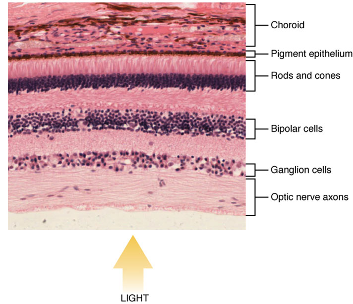
Nuclei of rods and cones The nuclei of rods and cones form a thick, densely packed layer within the retina, visible under the microscope at 800x magnification. This layer contains the genetic material and supports the metabolic activity essential for photoreceptor function.
Anatomy of Retinal Photoreceptors
The retina houses photoreceptors, and this microscope view highlights their cellular foundation at a detailed level. The image, provided by the Regents of University of Michigan Medical School, showcases the critical nuclear layer at 800x magnification.
- The nuclei of rods and cones represent the outer nuclear layer, where rod and cone cell bodies reside.
- Rods, more numerous, have nuclei that outnumber those of cones, reflecting their prevalence.
- Cones, though fewer, have nuclei concentrated in areas like the fovea for high-acuity vision.
- The dense packing of these nuclei supports efficient signal processing within the retina.
- Surrounding cells, such as Müller cells, provide structural support and nourishment.
- The micrograph’s 800x magnification reveals cellular details, aiding in anatomical study.
Physiology of Photoreceptor Nuclei
The nuclei of rods and cones are central to the photoreceptors’ ability to detect light and transmit signals. This microscopic perspective illustrates their role in sustaining visual function.
- The nuclei contain DNA, directing protein synthesis for photopigment production.
- Rod nuclei support the high sensitivity needed for low-light vision, with more cells per unit area.
- Cone nuclei, less dense, are tailored for color vision and detailed sight in bright conditions.
- Metabolic activity within these nuclei powers the phototransduction process.
- The outer nuclear layer connects to inner segments, where energy production occurs.
- Damage to this layer can disrupt the entire visual pathway, though this image shows healthy tissue.
Role of Rod Nuclei in Low-Light Vision
The nuclei of rods and cones include a significant population of rod nuclei, optimized for dim environments. Their structure supports the retina’s night vision capabilities.
- Rod nuclei are more abundant, enabling widespread light detection in low-light settings.
- These nuclei regulate rhodopsin synthesis, the pigment critical for dim-light sensitivity.
- The dense arrangement maximizes signal integration from rod photoreceptors.
- Rods lack color sensitivity, relying on a single photopigment type controlled by these nuclei.
- The outer nuclear layer’s thickness reflects rod prevalence, especially outside the fovea.
- Adaptation to darkness involves nuclear activity in rhodopsin regeneration.
Role of Cone Nuclei in Color Vision
The nuclei of rods and cones also encompass cone nuclei, designed for bright-light and color vision. Their organization supports high-resolution visual tasks.
- Cone nuclei are fewer but strategically located, particularly in the fovea’s center.
- These nuclei govern the production of opsins, pigments for red, green, and blue light detection.
- The sparser distribution allows for precise spatial resolution and color differentiation.
- Cone nuclei support faster photopigment renewal, enhancing adaptability to light changes.
- Their concentration in the fovea aligns with the retina’s sharpest vision area.
- Loss of cone nuclei function can lead to color vision deficits.
Microscopic Insights into Retinal Tissue
This 800x magnification micrograph, courtesy of the Regents of University of Michigan Medical School, offers a close-up of retinal tissue. The image emphasizes the cellular density and organization of photoreceptor nuclei.
- The nuclei of rods and cones appear as a distinct band, highlighting their layered structure.
- Staining techniques enhance the visibility of these nuclei, revealing their density.
- The micrograph shows the outer nuclear layer’s thickness, a key histological feature.
- Adjacent layers, like the outer plexiform layer, contain synaptic connections not visible here.
- The image supports research into retinal cell distribution and health.
- Variations in nuclear size and shape can indicate developmental or pathological changes.
Clinical Relevance of Photoreceptor Nuclei
Understanding the nuclei of rods and cones aids in diagnosing and managing retinal conditions. This micrograph serves as a reference for assessing normal retinal histology.
- Retinitis pigmentosa begins with rod nuclei degeneration, leading to night blindness.
- Age-related macular degeneration affects cone nuclei in the fovea, causing central vision loss.
- Thinning of the nuclei of rods and cones layer is an early sign of retinal disease.
- Genetic mutations impacting these nuclei can result in inherited retinal dystrophies.
- Optical coherence tomography measures nuclear layer thickness for diagnostic purposes.
- Treatments like gene therapy aim to preserve or restore nuclear function.
- Regular retinal exams monitor these nuclei for signs of deterioration.
In conclusion, the microscope view of photoreceptor nuclei offers a fascinating insight into the retina’s cellular foundation. This detailed image underscores the critical role of rods and cones in vision, providing a valuable resource for exploring the complexities of ocular health.

