Euglena is a fascinating mixotrophic protist that bridges the worlds of plants and animals, capable of both photosynthesis and heterotrophic feeding depending on environmental conditions. This unicellular organism, belonging to the supergroup Excavata, serves as an important model for studying photosynthesis, phototaxis, and metabolic flexibility in eukaryotes. The detailed diagram and microscopic image highlight its unique structural adaptations that allow survival in diverse aquatic habitats, offering valuable insights for students, researchers, and clinicians interested in protist biology and evolutionary cell processes.
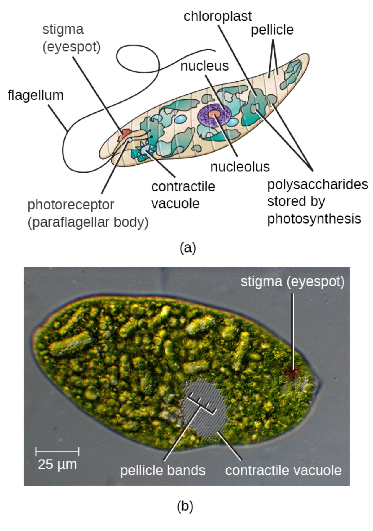
Stigma (eyespot) is the red-orange pigmented structure located near the anterior end. It acts as a shading device for the photoreceptor, enabling the organism to detect the direction of light and perform positive or negative phototaxis to optimize photosynthetic activity.
Flagellum is the long, whip-like structure extending from the anterior reservoir. It propels the cell through water using a characteristic helical motion, allowing Euglena to move toward optimal light or nutrient conditions.
Photoreceptor (paraflagellar body) is the light-sensitive swelling at the base of the flagellum. It works in conjunction with the stigma to sense light intensity and direction, triggering behavioral responses that position the cell for maximum photosynthesis.
Chloroplast is the green organelle responsible for photosynthesis. It contains chlorophyll and other pigments, enabling Euglena to produce energy from sunlight when light is available, while switching to heterotrophic nutrition in darkness.
Pellicle is the flexible proteinaceous strip layer beneath the plasma membrane. It provides structural support and allows the cell to change shape through euglenoid movement, also known as metaboly, which aids in locomotion and feeding.
Nucleus is the large central structure containing the genetic material. It controls cellular activities and is clearly visible in both the diagram and microscopic view, reflecting the typical eukaryotic organization of Euglena.
Nucleolus is the dense region within the nucleus involved in ribosome synthesis. Its prominent appearance highlights the high transcriptional activity required for the organism’s rapid growth and metabolic versatility.
Contractile vacuole is the clear, spherical organelle that regulates osmotic pressure. It collects excess water and expels it periodically, preventing the cell from bursting in hypotonic freshwater environments.
Polysaccharides stored by photosynthesis refers to the paramylon granules produced and stored as energy reserves from photosynthetic activity. These carbohydrate stores allow Euglena to survive periods without light by switching to heterotrophic metabolism.
Pellicle bands are the visible helical strips of the pellicle seen in the microscopic image. They give Euglena its characteristic striated appearance and enable the flexible shape changes used during metaboly movement.
Biological Versatility of Euglena
Euglena exemplifies mixotrophy, the ability to combine autotrophy through photosynthesis and heterotrophy by absorbing organic compounds. In bright light, chloroplasts dominate energy production, while in darkness or nutrient-rich conditions, the organism ingests bacteria or organic matter through the cytostome. This metabolic flexibility provides a survival advantage in fluctuating aquatic environments and makes Euglena an excellent model for studying the evolutionary transition between plant-like and animal-like nutrition.
Phototaxis and Sensory Systems
The combination of stigma and photoreceptor allows Euglena to perform sophisticated phototaxis. The eyespot shades the photoreceptor as the cell rotates during swimming, creating a periodic signal that the organism uses to steer toward or away from light. This system demonstrates how a single cell can integrate sensory information and generate directed behavioral responses without a nervous system.
Structural Adaptations for Survival
The pellicle provides both protection and flexibility, allowing Euglena to squeeze through narrow spaces and perform euglenoid movement when flagellar propulsion is insufficient. The contractile vacuole maintains osmotic balance, while paramylon granules serve as energy reserves. These adaptations enable Euglena to thrive in diverse habitats ranging from freshwater ponds to temporary pools with varying light and nutrient availability.
- Flagellar movement enables rapid swimming toward light sources.
- Pellicle bands allow shape changes for crawling or squeezing.
- Contractile vacuole prevents cellular swelling in freshwater.
Such features highlight the sophisticated engineering present in unicellular eukaryotes.
Euglena as a Model Organism in Research
Euglena has been extensively used in laboratory studies of photosynthesis, circadian rhythms, and flagellar biology. Its chloroplasts are similar to those of higher plants, making it useful for investigating photosynthetic mechanisms. The organism is also employed in toxicity testing and environmental monitoring because of its sensitivity to pollutants and ease of cultivation. Modern genetic tools have further expanded its utility in synthetic biology and metabolic engineering.
Ecological and Educational Importance
In nature, Euglena contributes to primary production in aquatic ecosystems and serves as food for small invertebrates. In education, its large size and visible organelles make it ideal for microscopy labs, allowing students to observe living cells performing photosynthesis, movement, and osmoregulation in real time. The clear distinction between chloroplast-rich green cells and etiolated forms in darkness beautifully demonstrates metabolic switching.
Clinical and Applied Relevance
While Euglena itself is generally non-pathogenic, its study contributes to broader understanding of protist biology relevant to parasitic excavates such as trypanosomes. Research on its photoreceptor and flagellar systems informs fields ranging from bioengineering to environmental science. Additionally, Euglena biomass is explored for nutritional supplements and biofuel production due to its high protein and paramylon content.
Conclusion: The Remarkable Adaptability of Euglena
The structure and microscopic view of Euglena reveal a highly adapted unicellular organism capable of thriving through both photosynthetic and heterotrophic pathways. From the light-sensing stigma and flagellum to the flexible pellicle and energy-storing polysaccharides, every labeled feature supports its success in variable environments. As a model organism, Euglena continues to provide insights into eukaryotic evolution, cellular behavior, and practical applications in biotechnology and education, underscoring the enduring scientific value of this versatile protist.

