Understanding the early stages of human heart formation provides crucial insights into how this vital organ evolves from a simple tube into a complex four-chambered structure capable of sustaining life. At 28 days of gestation, significant partitioning begins, marking a pivotal phase in embryonic cardiovascular development where septa and cushions form to separate the atria and ventricles.
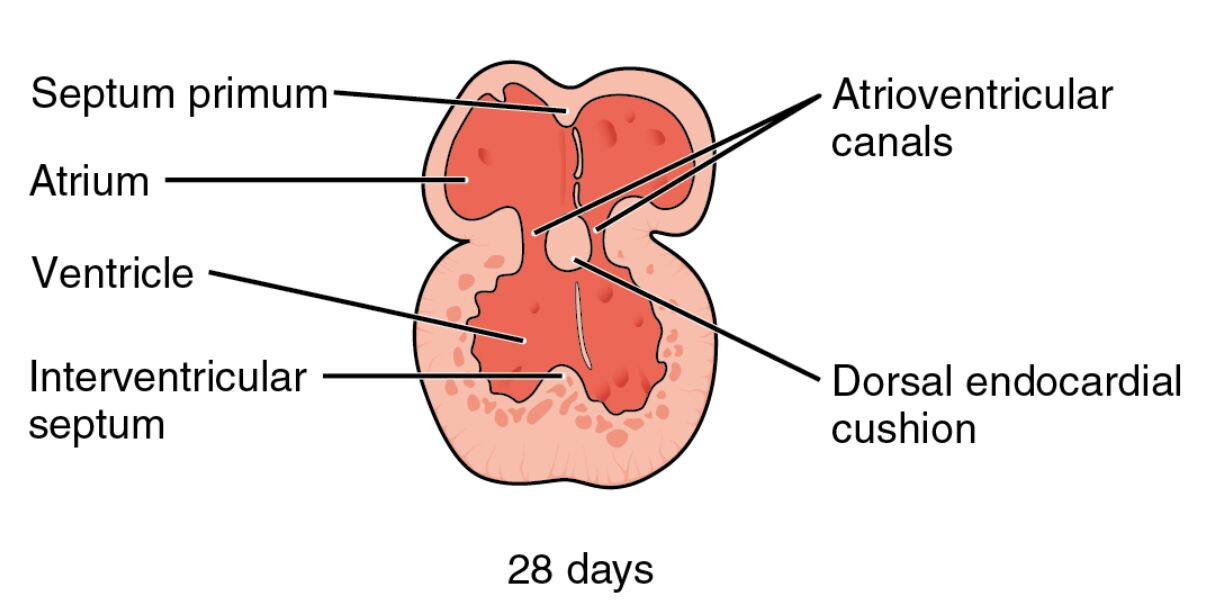
Septum primum The septum primum is a thin, crescent-shaped membrane that grows downward from the roof of the primitive atrium toward the endocardial cushions. This structure initiates the division of the single atrial chamber into right and left atria, ensuring proper separation of oxygenated and deoxygenated blood flows later in development. As it extends, it leaves an opening called the ostium primum, which allows blood to pass between the atria temporarily.
Atrium The atrium represents the upper chamber of the developing heart, initially as a single cavity that will eventually divide into two separate atria. It receives blood from the venous system and plays a key role in the embryonic circulation by directing flow toward the ventricle. During this stage, the atrium is expanding and beginning to partition, setting the foundation for efficient blood pumping in the mature heart.
Ventricle The ventricle is the lower chamber in the embryonic heart, starting as a unified space that will split into right and left ventricles. It is responsible for propelling blood into the arterial system, and at 28 days, muscular ridges are forming to aid in its division. This early ventricular structure ensures that the heart can handle increasing circulatory demands as the embryo grows.
Interventricular septum The interventricular septum is a muscular partition that grows upward from the floor of the primitive ventricle, contributing to the separation of the ventricular chambers. It fuses with other septa to create distinct right and left ventricles, preventing the mixing of blood streams. This septum’s development is essential for the heart’s ability to pump blood to the lungs and body separately in postnatal life.
Atrioventricular canals The atrioventricular canals are the passageways connecting the atrium to the ventricle, which will later become guarded by valves. At this stage, they allow blood to flow from the upper to the lower chambers and are flanked by endocardial cushions that will form the atrioventricular valves. Proper formation of these canals is critical to prevent congenital defects like atrioventricular septal abnormalities.
Dorsal endocardial cushion The dorsal endocardial cushion is one of the swellings in the atrioventricular canal, composed of mesenchymal tissue that protrudes into the canal. It merges with its ventral counterpart to divide the canal into right and left channels, eventually contributing to the formation of the mitral and tricuspid valves. This cushion’s role is vital in ensuring valvular integrity and proper heart function.
The Importance of Early Heart Partitioning
Early heart partitioning is a meticulously orchestrated process involving cellular migration, proliferation, and fusion to create functional chambers. This stage at 28 days highlights the transition from a primitive heart tube to a more advanced structure, influenced by genetic and environmental factors.
- Partitioning begins with the formation of septa that divide the heart’s interior spaces, preventing inefficient blood mixing.
- Key signaling pathways, such as those involving transforming growth factor-beta (TGF-β), regulate the growth of structures like the septum primum.
- Disruptions in this phase can lead to congenital heart defects, emphasizing the need for precise developmental timing.
- The embryonic heart beats by this point, supporting nutrient distribution throughout the growing fetus.
- Imaging techniques like ultrasound can detect these early structures in prenatal screenings.
Key Structures in 28-Day Embryonic Heart
At 28 days, the heart’s anatomy is rapidly evolving, with visible septa and cushions indicating progress toward chamber separation. This diagram captures a transverse section, illustrating how these elements interact to form the four-chambered heart.
- The septum primum extends inferiorly, approaching the endocardial cushions to close off the atrial space.
- The single atrium is poised for division, with blood entering via sinus venosus connections.
- The ventricle shows initial muscular trabeculations, precursors to the adult ventricular walls.
- The interventricular septum rises from the apex, eventually meeting the atrial septum.
- Atrioventricular canals are narrowing due to cushion growth, setting up valve formation.
- The dorsal endocardial cushion, along with others, derives from neural crest cells and endocardial epithelium.
Physiological Implications of Heart Development
The physiology of the developing heart adapts to the embryo’s needs, with shunts allowing oxygenated blood to bypass non-functional lungs. At this stage, the heart’s partitioning supports efficient circulation within the yolk sac and placental systems.
- Blood flow through the atrioventricular canals is unidirectional, driven by peristaltic contractions.
- Oxygenation occurs via diffusion from maternal blood, not pulmonary means.
- Hormonal influences, such as thyroid hormones like T3 and T4, may modulate cardiac cell differentiation indirectly through maternal circulation.
- The embryonic heartbeat rate is around 100-120 beats per minute, increasing with development.
- Nutrient delivery to cardiac tissues ensures continued growth and septation.
Genetic and Molecular Basis
Genetic factors underpin the structural changes seen in this diagram, with transcription factors guiding cell fate. Molecular interactions ensure that septa form correctly, involving genes like NKX2-5 and GATA4.
- Mutations in these genes can disrupt septum formation, leading to atrial or ventricular septal defects.
- Epithelial-mesenchymal transition (EMT) is crucial for endocardial cushion development.
- Signaling from the cardiac neural crest contributes to outflow tract and septal alignment.
- Extracellular matrix components, like hyaluronic acid, provide scaffolding for migrating cells.
- Epigenetic modifications regulate the timing of gene expression during partitioning.
Comparative Embryology Across Species
Comparing human heart development to other vertebrates reveals conserved mechanisms, though timelines differ. In mammals, the 28-day stage in humans corresponds roughly to similar phases in mice or chicks, offering models for research.
- Avian models show similar septum primum growth, aiding in experimental manipulations.
- Mammalian similarities ensure translational relevance for congenital defect studies.
- Evolutionary adaptations have refined partitioning for efficient four-chamber circulation.
- Differences in cushion formation can explain species-specific heart morphologies.
- Research using zebrafish highlights rapid heart development for genetic screening.
Clinical Relevance and Prenatal Monitoring
Clinically, understanding this stage aids in diagnosing early anomalies via advanced imaging. Prenatal care focuses on detecting deviations that could impact fetal viability.
- Echocardiography can visualize septa and cushions by the first trimester.
- Genetic testing identifies risks for defects like atrioventricular canal malformations.
- Maternal health factors, including nutrition, influence developmental outcomes.
- Interventions, such as folic acid supplementation, support normal heart formation.
- Long-term, early detection improves surgical planning for congenital issues.
Future Research Directions
Ongoing research explores regenerative medicine inspired by embryonic processes. Stem cell studies aim to recapitulate partitioning for heart repair.
- Induced pluripotent stem cells (iPSCs) can model 28-day-like structures in vitro.
- CRISPR gene editing targets developmental genes for defect correction.
- Bioinformatics analyzes genomic data from embryonic tissues.
- 3D bioprinting seeks to replicate septal growth for transplants.
- Collaborative efforts integrate embryology with cardiology for better therapies.
The intricate process depicted in this 28-day embryonic heart diagram underscores the marvel of human development, where precise cellular events lay the groundwork for lifelong cardiovascular health. By appreciating these early stages, we gain deeper respect for the organ that sustains us from the womb onward, highlighting the importance of continued study in embryology to advance medical knowledge and interventions.

