The development of the human heart progresses notably by day 23, revealing the formation of key structures such as the truncus arteriosus, bulbus cordis, ventricle, atrium, and sinus venosus within the primitive heart tube. This image captures the heart’s early looping and segmentation, a pivotal stage where the circulatory system begins to support the embryo’s growth through initial contractions.
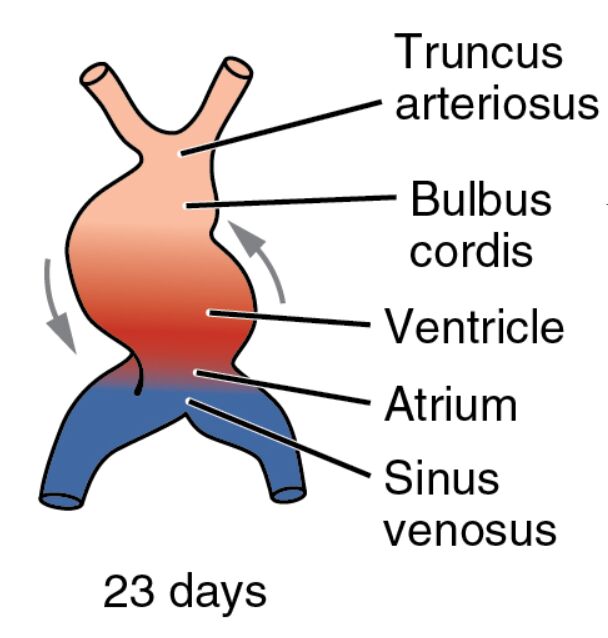
Truncus arteriosus Truncus arteriosus forms the outflow tract, directing blood from the heart to the developing arterial system, located at the cranial end of the heart tube. This structure will undergo septation to create the aorta and pulmonary artery, establishing dual circulation in later stages.
Bulbus cordis Bulbus cordis is a dilated region near the truncus arteriosus, contributing to the future right ventricle and outflow tracts. Its development involves significant remodeling to align with the heart’s eventual chambered structure.
Ventricle Ventricle serves as the primary pumping chamber, propelling blood through the bulbus cordis and truncus arteriosus, centrally located in the looping heart tube. This region will divide into the left and right ventricles, each adapted for specific circulatory roles.
Atrium Atrium acts as the receiving chamber for blood returning via the sinus venosus, positioned caudally in the heart tube. It will expand and differentiate into the left and right atria, incorporating venous inputs over time.
Sinus venosus Sinus venosus is the venous inflow tract, collecting deoxygenated blood from the vitelline, umbilical, and common cardinal veins at the heart’s caudal end. This structure will contribute to the right atrium and the sinoatrial node, initiating heartbeats in the mature organ.
23 days 23 days marks the gestational age, corresponding to Carnegie stage 10-11, when the heart tube completes its initial looping and begins functional activity. The embryo, about 2.5-3.5 mm long, relies on this early heart development for nutrient and oxygen distribution.
The Significance of Heart Looping and Early Function
This stage represents a critical transition in heart development, with looping shaping its structure. The onset of contractions marks the beginning of a functional circulatory system.
- Looping Mechanism: The heart tube’s S-shaped loop forms due to differential growth, guided by NKX2-5 and Pitx2 signaling. This process ensures proper alignment of future chambers.
- Contraction Onset: Rhythmic contractions start, driven by the myocardial layer’s calcium signaling. These beats support early circulation from the yolk sac.
- Segmentation Initiation: The heart tube begins segmenting into truncus arteriosus, bulbus cordis, ventricle, atrium, and sinus venosus. This specialization lays the groundwork for chamber formation.
Anatomical Features of the 23-Day Embryo
The image illustrates the heart tube’s S-shaped loop, with arrows indicating blood flow direction. This visual representation highlights the dynamic changes in cardiac anatomy at this stage.
The embryo’s folding continues to position the heart within the thoracic cavity, offering protection. Color gradients reflect the transition from venous to arterial blood flow, aiding in anatomical study.
- Truncus Arteriosus Structure: This outflow tract connects to the aortic sac, with an endothelial lining forming its inner layer. Neural crest cells will later migrate to initiate septation.
- Bulbus Cordis Anatomy: Located between the ventricle and truncus, this region thickens its myocardial walls. It will contribute to the right ventricle and conus arteriosus.
- Ventricle Characteristics: The ventricle’s walls begin to thicken, indicating its pumping role. Trabeculae start forming to enhance contraction strength.
- Atrium Details: The atrium receives blood from the sinus venosus, with early septation beginning. This chamber will grow to accommodate venous return.
- Sinus Venosus Features: This venous collector merges with the atrium, with its horns receiving blood from major veins. It will form part of the right atrium and pacemaker region.
Developmental Milestones at 23 Days
This phase brings notable progress, with the heart’s looping and initial contractions driving circulatory development. The coordination with other systems reflects the embryo’s complexity.
The neural tube approaches closure, and somites form along the body axis, paralleling cardiac maturation. These milestones ensure balanced embryonic growth.
- Looping Progression: The S-shape loop continues to refine, guided by left-right asymmetry signals. This alignment is crucial for future chamber positioning.
- Cellular Migration: Cardiac neural crest cells begin migrating to the outflow tract, aiding truncus arteriosus septation. This prevents congenital outflow issues.
- Vascular Connections: The sinus venosus links to vitelline and umbilical veins, forming a rudimentary loop. This network supports nutrient delivery from the yolk sac.
- Genetic Influence: Genes like GATA4 enhance myocardial differentiation, guiding chamber development. Mutations can lead to atrial septal defects.
Physiological Functions of the Looping Heart
The looping heart starts performing essential physiological roles, marking the shift to active circulation. Its early contractions meet the embryo’s growing demands.
The heart’s rudimentary pump operates with low pressure, relying on the yolk sac for oxygenation. This efficiency sets the stage for fetal circulatory adaptations.
- Pumping Action: The ventricle’s contractions propel blood through the bulbus cordis and truncus arteriosus. This flow supports embryonic metabolic needs.
- Blood Flow Direction: Deoxygenated blood enters via the sinus venosus, while blood exits through the truncus arteriosus. This pattern anticipates dual circulation.
- Oxygen Supply: Primitive erythrocytes from blood islands carry limited oxygen, supplemented by the yolk sac. This sustains the embryo until placental circulation develops.
- Pressure Gradients: The looped structure creates pressure differences, guiding vascular remodeling. These gradients influence arterial and venous formation.
Clinical and Research Implications
The 23-day embryo provides valuable insights for medical practice and research. The heart’s looping and early function are key areas for studying congenital conditions.
Advanced imaging and stem cell research deepen our understanding of this stage. These tools offer potential for early diagnosis and innovative treatments.
- Congenital Defects: Looping errors can cause dextrocardia, detectable via ultrasound. Early detection improves management strategies.
- Regenerative Medicine: Stem cells can be induced to form looped heart structures. This approach supports cardiac repair efforts.
- Animal Models: Chick and mouse embryos show similar looping patterns. These models validate human developmental studies.
- Therapeutic Targets: Modulating VEGF pathways may enhance vascular growth in defects. Gene editing explores correcting cardiac gene mutations.
In conclusion, this image of the 23-day embryo highlights the heart’s looping and segmentation into key regions, a vital step in cardiovascular development. This early structuring supports embryonic life and provides a foundation for understanding and addressing heart-related health challenges.

