The early stages of human embryonic development reveal fascinating insights into how the cardiovascular system begins to form, with the heart emerging as one of the first functional organs. At just 18 days post-fertilization, the embryo displays critical structures like the cardiogenic area and primitive blood vessels, setting the foundation for a complex circulatory network that will support growth throughout gestation.
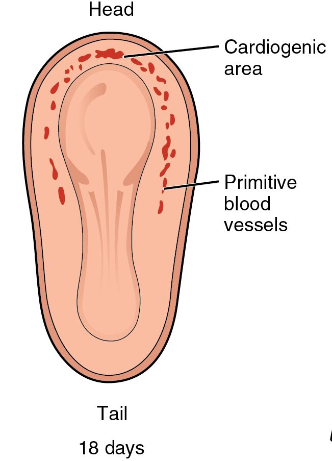
Head The head region of the embryo at this stage is the anterior end, where rapid cellular differentiation is occurring to form neural and facial structures. This area is crucial as it houses the developing brain and sensory organs, influencing overall embryonic patterning.
Tail The tail represents the posterior end of the embryo, which will eventually regress as development progresses. It plays a role in establishing the body axis and contains cells that contribute to the formation of the spinal cord and other caudal structures.
Cardiogenic area The cardiogenic area is a specialized region of mesoderm located near the head, destined to give rise to the heart tube. This zone is where cardiac progenitor cells aggregate and begin to differentiate, marking the onset of cardiogenesis essential for embryonic survival.
Primitive blood vessels Primitive blood vessels appear as early vascular formations scattered throughout the embryo, facilitating nutrient and oxygen transport before a fully formed circulatory system develops. These structures arise from angioblasts and will coalesce to form more mature vessels, supporting the growing embryo’s metabolic needs.
18 days The notation of 18 days indicates the gestational age of the embryo depicted, corresponding to Carnegie stage 8 in human development. At this point, key milestones like gastrulation have occurred, paving the way for organogenesis, including the initial steps in heart formation.
The Significance of Early Cardiovascular Formation
Understanding the timeline of embryonic heart development provides a window into the intricate processes that ensure proper organ function from the outset. This phase is marked by cellular migrations and inductions that lay the groundwork for lifelong health.
The heart begins to form remarkably early in pregnancy, driven by genetic and environmental cues that orchestrate cell fate decisions. Disruptions at this stage can lead to congenital anomalies, highlighting the importance of studying these initial structures.
- Embryonic Layers Involved: The process starts with the three primary germ layers—ectoderm, mesoderm, and endoderm—established during gastrulation. The mesoderm, in particular, gives rise to the cardiogenic mesoderm, which is visible in the image as the cardiogenic area.
- Role of Signaling Pathways: Pathways like Wnt, BMP, and FGF signaling regulate the specification of cardiac cells. These molecular signals ensure that progenitor cells in the cardiogenic area commit to a cardiac lineage, preventing inappropriate differentiation.
- Integration with Other Systems: Even at 18 days, the developing heart interacts with the nascent nervous system and yolk sac, emphasizing the interconnected nature of embryonic organogenesis.
Detailed Anatomy of the 18-Day Embryo
Visual representations like this illustration capture the embryo’s pear-like shape, with distinct head and tail regions orienting the viewer. The red markings highlight areas of active vasculogenesis, offering a clear depiction of primitive structures.
At this stage, the embryo measures about 1-1.5 mm in length, curled in a C-shape due to the folding of the body walls. This configuration protects vital developing organs and facilitates nutrient exchange via the yolk sac.
- Cardiogenic Area in Depth: Positioned cranially, this region consists of splanchnic mesoderm that will fold to form the endocardial tubes. These tubes fuse to create the primitive heart tube, which begins beating around day 21-22.
- Formation of Primitive Blood Vessels: These vessels emerge through vasculogenesis, where endothelial cells differentiate from mesodermal precursors called angioblasts. They form a network that initially lacks connection but soon links up to enable circulation.
- Embryonic Folding and Its Impact: Lateral and longitudinal folding brings the cardiogenic area into the thoracic region, a critical morphogenetic event. This movement ensures the heart is properly positioned relative to the developing pharyngeal arches.
- Blood Islands and Hematopoiesis: Scattered red areas in the image likely represent blood islands, sites where primitive erythrocytes form. This early blood production is vital for oxygen delivery before the placenta fully develops.
Developmental Milestones Around Day 18
Milestones in embryonic development are tightly regulated, with each day bringing new advancements in structure and function. By day 18, the groundwork for major organs is laid, setting the stage for rapid growth in subsequent weeks.
The notochord, a rod-like structure, induces neural tube formation, while somites begin to segment along the body axis. These events occur in tandem with cardiovascular initiation, illustrating the synchronized nature of embryogenesis.
- Preceding Events: Prior to day 18, implantation and gastrulation establish the basic body plan. The primitive streak forms, allowing cells to ingress and form mesoderm, including cardiac precursors.
- Subsequent Developments: Following this stage, the heart tube elongates and loops, forming chambers like the bulbus cordis and ventricle. By week 4, septation begins, dividing the heart into left and right sides.
- Genetic Regulation: Genes such as NKX2-5 and GATA4 are expressed in the cardiogenic area, acting as transcription factors for cardiac differentiation. Mutations in these can lead to defects like atrial septal defects.
- Environmental Influences: Maternal factors, including nutrition and exposure to teratogens, can affect this delicate phase. For instance, folic acid deficiency may impair neural tube closure, indirectly impacting heart development.
Physiological Implications of Early Heart Structures
The physiology of the embryonic heart differs from the adult, relying on diffusion initially before active pumping starts. These primitive structures ensure survival in the low-oxygen uterine environment.
Blood flow in the early embryo is sluggish, driven by peristaltic-like contractions once the heart tube forms. This sets up gradients for nutrient distribution and waste removal.
- Oxygenation Mechanisms: Primitive blood vessels connect to the yolk sac, which acts as a temporary respiratory organ. Vitelline circulation provides oxygen until placental development.
- Hormonal and Growth Factors: Factors like VEGF promote angiogenesis in primitive vessels. This growth factor ensures vessel sprouting and maturation.
- Transition to Fetal Circulation: From these beginnings, shunts like the ductus arteriosus develop to bypass non-functional lungs. Understanding this evolution aids in diagnosing fetal anomalies via ultrasound.
- Comparative Embryology: Similar patterns occur in other vertebrates, with the cardiogenic area conserved across species. This homology provides insights into evolutionary biology.
Clinical Relevance and Research Advances
Insights from embryonic images like this inform clinical practices in reproductive medicine and cardiology. They help in identifying critical windows for intervention in developmental disorders.
Advances in imaging techniques, such as high-resolution ultrasound, allow monitoring of these stages in vivo. Research using stem cell models replicates cardiogenesis for therapeutic purposes.
- Congenital Heart Disease Links: Many defects trace back to errors in the cardiogenic area, such as tetralogy of Fallot. Early detection through genetic screening can improve outcomes.
- Stem Cell Therapies: Induced pluripotent stem cells can be directed to form cardiac tissue, mimicking the primitive structures shown. This holds promise for regenerative medicine.
- Ethical Considerations in Research: Studying human embryos raises questions about sourcing and consent. Animal models often supplement to bridge knowledge gaps.
- Future Directions: CRISPR gene editing targets cardiac genes to prevent defects. Integrating AI in analyzing embryonic images accelerates discoveries.
In summary, this depiction of the 18-day embryo underscores the miraculous precision of human development, where simple structures like the cardiogenic area and primitive blood vessels evolve into a vital organ system. Continued exploration of these early phases not only deepens our understanding of biology but also enhances medical strategies to support healthy beginnings.

