Embryonic folding is a critical and complex process in early human development that transforms a flat, trilaminar embryonic disc into a cylindrical, three-dimensional structure. This fundamental re-shaping establishes the basic body plan and facilitates the formation of crucial internal organs, most notably the primitive gut tube. The provided diagram illustrates the dynamic movements of embryonic folding, showing how the various germ layers contribute to this remarkable metamorphosis. Understanding this process is essential for comprehending the foundational development of all organ systems and the potential origins of congenital anomalies.
Key Components and Stages of Embryonic Folding
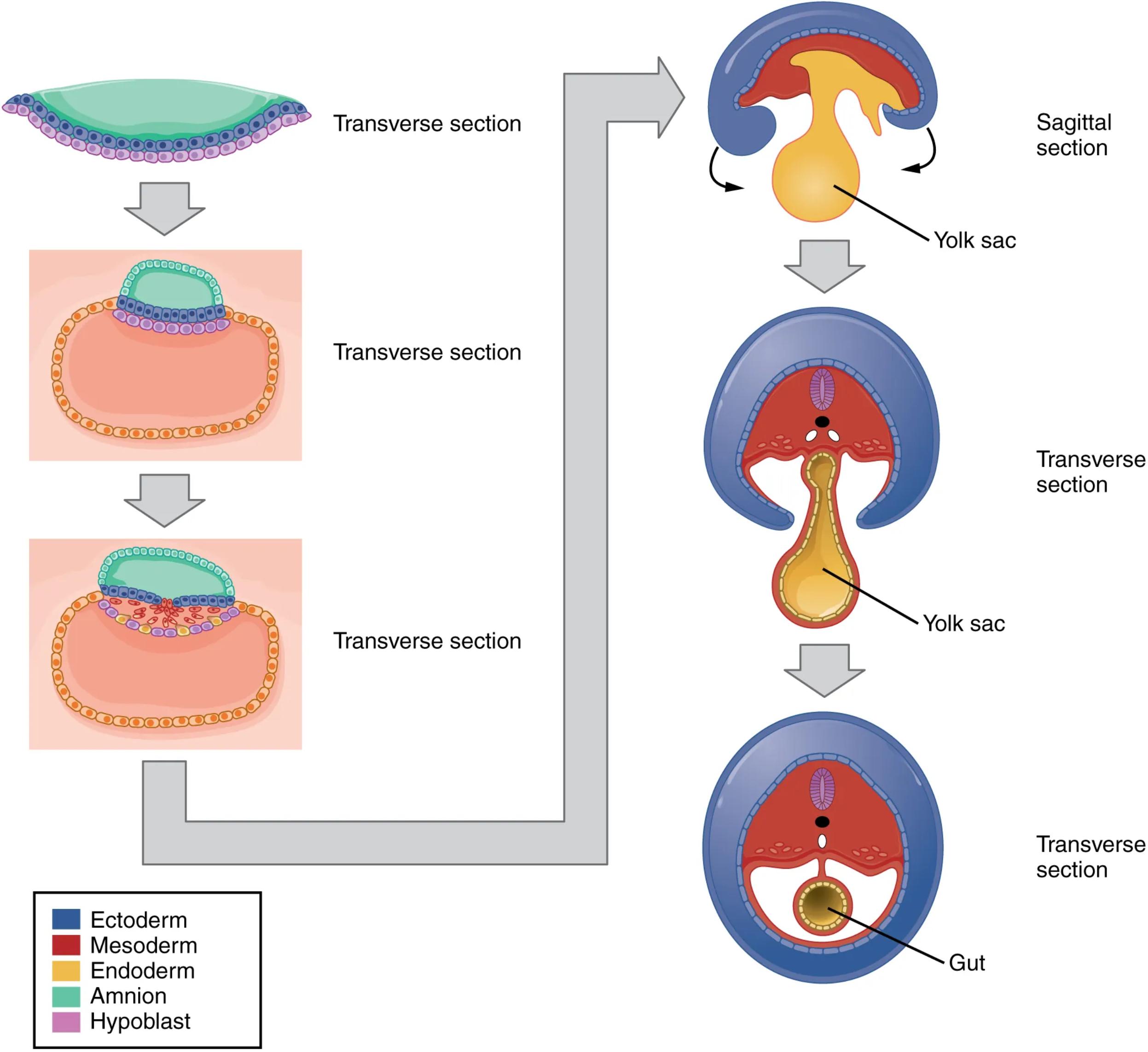
Ectoderm: Represented in blue, the ectoderm is the outermost of the three primary germ layers. During folding, it will largely form the external surface of the embryo, giving rise to the epidermis and the nervous system.
Mesoderm: Shown in red, the mesoderm is the middle germ layer. It plays a crucial role in forming connective tissues, muscle, bone, and the cardiovascular system, and its lateral plates are instrumental in the folding process.
Endoderm: Depicted in orange, the endoderm is the innermost germ layer. It primarily forms the lining of the gastrointestinal and respiratory tracts, as well as associated glandular organs.
Amnion: Indicated in green, the amnion is a fetal membrane that encloses the embryo, forming the amniotic cavity filled with amniotic fluid. During folding, the amnion expands to surround the entire embryo.
Hypoblast: Represented in pink, the hypoblast is an early embryonic cell layer that contributes to the extraembryonic membranes, particularly the yolk sac. It is foundational in establishing the primitive streak.
Transverse section: These diagrams illustrate the embryonic disc cut horizontally, showing the germ layers and surrounding structures from a cross-sectional perspective. They highlight the lateral folding of the embryo.
Sagittal section: This diagram displays the embryo cut vertically along the midline, revealing the cranial (head) and caudal (tail) folding. It effectively shows how the head and tail ends curve ventrally.
Yolk sac: This extraembryonic membrane, shown in yellow, is initially a prominent structure continuous with the endoderm. During folding, portions of the yolk sac are incorporated into the embryo to form the primitive gut.
Gut: The primitive gut tube, formed from the incorporated endoderm of the yolk sac, is the precursor to the entire gastrointestinal tract. This tube develops as a result of the inward folding of the embryonic disc.
The Dynamics of Embryonic Folding
Embryonic folding occurs simultaneously in two planes: cephalocaudal (head-to-tail) and lateral (side-to-side). These coordinated movements transform the flat trilaminar disc into a C-shaped embryo. The cephalocaudal folding is driven primarily by the differential growth of the neural tube and the adjacent somites, causing the head and tail regions to curl ventrally towards the midline. This process brings the developing heart (initially cranial) and the cloacal membrane (caudal) into their definitive ventral positions.
Concurrently, lateral folding involves the inward bending of the two lateral edges of the embryonic disc. This movement is largely influenced by the rapid growth of the somites and the expanding amniotic cavity. As the lateral folds meet and fuse at the ventral midline, they progressively enclose a portion of the yolk sac, forming the primitive gut tube. This fusion also brings the two lateral body walls together, establishing a continuous outer body surface derived from the ectoderm.
- The precise timing and coordination of these folding events are paramount for normal development; disruptions can lead to significant birth defects, such as omphalocele or gastroschisis, where abdominal contents protrude externally due to incomplete ventral body wall closure.
Ultimately, embryonic folding creates the distinct tubular body plan, separating the internal organs from the external environment and laying down the foundations for all major organ systems. The remaining parts of the yolk sac are pinched off and become the vitelline duct, which typically regresses.
Conclusion
Embryonic folding is a fundamental event in human embryogenesis, marking the transition from a flat, two-dimensional disc to a three-dimensional embryo with a recognizable body shape. This diagram effectively illustrates the profound changes in morphology, showing how the ectoderm, mesoderm, and endoderm are repositioned to form the basic body plan, including the crucial gut tube. Understanding these intricate folding processes is essential for grasping the subsequent development of organ systems and for recognizing the embryological basis of various congenital malformations.

