The elbow joint is a complex hinge joint that facilitates flexion and extension of the forearm, supported by key ligaments like the ulnar and radial collateral ligaments, and the annular ligament at the proximal radioulnar joint. This structure allows for precise movements while ensuring stability, making it essential for activities like lifting and throwing. This article explores the anatomical structure of the elbow joint, its physical functions, and its role in upper limb mobility, providing a comprehensive understanding of its components and significance.
Labeled Parts of the Elbow Joint Structure
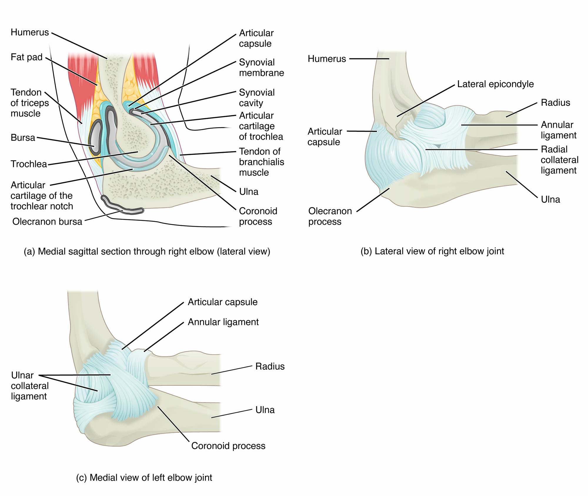
Humerus
The humerus is the upper arm bone that forms the proximal part of the elbow joint, articulating with the radius and ulna at its distal end. Its trochlea and capitulum provide smooth surfaces for the hinge motion of the elbow and the rotation of the radius, respectively.
Ulna
The ulna is one of the two forearm bones, located on the medial side, and articulates with the humerus at the elbow joint through its trochlear notch. It plays a primary role in the hinge motion of the elbow, allowing for flexion and extension of the forearm.
Radius
The radius is the lateral forearm bone that articulates with the humerus at the elbow joint and rotates at the proximal radioulnar joint. Its head is supported by the annular ligament, enabling rotational movements like pronation and supination of the forearm.
Ulnar Collateral Ligament
The ulnar collateral ligament is a strong band of fibrous tissue on the medial side of the elbow, connecting the humerus to the ulna. It provides stability by resisting valgus stress, protecting the joint during movements like throwing or lifting.
Radial Collateral Ligament
The radial collateral ligament is located on the lateral side of the elbow, connecting the humerus to the radius and ulna. It stabilizes the joint by resisting varus stress, ensuring the elbow remains aligned during forearm motion.
Annular Ligament
The annular ligament is a ring-shaped band of fibrous tissue that encircles the head of the radius at the proximal radioulnar joint, a pivot joint. It secures the radius to the ulna, allowing for smooth rotation of the forearm during movements like turning the palm up or down.
Anatomical Structure of the Elbow Joint
Components of the Elbow Joint
The elbow joint is a synovial hinge joint composed of three articulations that work together to allow for both hinge and pivot movements. Its structure is designed to balance mobility with stability, supported by a network of ligaments and bones.
- The humerus articulates with the ulna at the humeroulnar joint and with the radius at the humeroradial joint, forming the primary hinge of the elbow.
- The ulna’s trochlear notch fits into the humerus’ trochlea, enabling the elbow’s flexion and extension, such as during bending or straightening the arm.
- The radius’ head articulates with the humerus’ capitulum, contributing to both the hinge motion of the elbow and the rotational motion at the proximal radioulnar joint.
- The ulnar collateral ligament and radial collateral ligament reinforce the joint medially and laterally, preventing excessive sideways movement.
- The annular ligament stabilizes the proximal radioulnar joint, a pivot joint that allows the radius to rotate over the ulna during forearm pronation and supination.
Supporting Ligaments and Joint Capsule
The ligaments and joint capsule of the elbow joint provide essential support, ensuring stability while allowing for a wide range of movements. These structures are critical for maintaining joint integrity under mechanical stress.
- The ulnar collateral ligament, with its three bands (anterior, posterior, and oblique), is particularly strong and crucial for resisting valgus forces during activities like pitching.
- The radial collateral ligament, a fan-shaped structure, prevents varus stress, maintaining lateral stability during forearm movements.
- The annular ligament forms a complete ring around the radial head, securing it against the ulna while allowing smooth rotation at the proximal radioulnar joint.
- The joint capsule, a fibrous sleeve enclosing the elbow joint, is lined with a synovial membrane that produces synovial fluid for lubrication.
- The capsule is reinforced by the collateral ligaments, ensuring the joint remains stable while permitting flexion, extension, and rotation.
Physical Introduction to the Elbow Joint
Biomechanical Functions of the Elbow Joint
The elbow joint’s hinge and pivot design enables precise movements of the forearm, making it essential for upper limb functionality. Its biomechanical properties ensure efficient motion while maintaining stability during dynamic activities.
- The humeroulnar joint acts as a hinge, allowing flexion and extension, which are critical for tasks like lifting objects or pushing.
- The humeroradial joint contributes to both hinge motion and forearm rotation, supporting movements like turning a doorknob or using a screwdriver.
- The proximal radioulnar joint, stabilized by the annular ligament, functions as a pivot joint, enabling pronation and supination of the forearm.
- The ulnar and radial collateral ligaments distribute mechanical stress across the joint, preventing injury during high-force activities like throwing.
- Synovial fluid within the joint capsule reduces friction between the articulating surfaces, ensuring smooth and pain-free movement.
Range of Motion and Movement Types
The elbow joint supports a specific range of motion that facilitates a variety of upper limb activities, with its hinge and pivot components working in tandem. This dual functionality makes the elbow a versatile joint for daily tasks.
- Flexion, the bending of the elbow, typically reaches about 145 degrees, allowing the forearm to approach the upper arm during actions like eating.
- Extension, the straightening of the elbow, returns the forearm to its anatomical position, essential for pushing or reaching movements.
- Pronation, the rotation of the radius over the ulna, turns the palm downward, useful for activities like typing or pouring water.
- Supination, the opposite rotation, turns the palm upward, enabling actions like holding a bowl or turning a key.
- The combination of hinge and pivot motions allows the elbow to contribute to complex movements, such as those required in sports or manual labor.
Clinical Insights: Elbow Joint Conditions
Common Disorders of the Elbow Joint
The elbow joint, despite its robust structure, is prone to various conditions that can affect its function and cause discomfort. Understanding these disorders is key to effective diagnosis and management.
- Tennis elbow (lateral epicondylitis) involves inflammation of the tendons on the lateral side of the elbow, often due to repetitive wrist extension, causing pain near the radial collateral ligament.
- Golfer’s elbow (medial epicondylitis) affects the medial side, near the ulnar collateral ligament, due to repetitive wrist flexion, leading to pain and tenderness.
- Elbow dislocations, often involving the humeroulnar joint, can occur due to trauma, potentially damaging the collateral ligaments and requiring immediate realignment.
- Ulnar collateral ligament injuries, common in throwing athletes, result from repetitive valgus stress, leading to instability and sometimes requiring surgical reconstruction.
- Radial head subluxation, also known as “nursemaid’s elbow,” occurs when the annular ligament slips over the radial head, typically in young children, causing pain and limited motion.
Prevention and Management of Elbow Joint Issues
Maintaining elbow joint health is essential for preserving upper limb function and preventing long-term complications. Proactive strategies and targeted interventions can help manage and prevent elbow-related problems.
- Strengthening the forearm muscles through exercises like wrist curls can reduce strain on the tendons, preventing conditions like tennis elbow or golfer’s elbow.
- Proper warm-up and stretching before sports or repetitive activities can minimize the risk of ligament injuries, such as ulnar collateral ligament tears.
- Avoiding sudden, forceful movements in young children can prevent radial head subluxation, ensuring the annular ligament remains intact.
- Rest, ice, and anti-inflammatory medications can alleviate symptoms of tennis elbow or golfer’s elbow, while physical therapy can improve tendon healing.
- In severe cases, surgical interventions like ulnar collateral ligament reconstruction or radial head realignment may be necessary to restore joint stability.
Conclusion
The elbow joint, with its hinge and pivot components, including the humerus, ulna, radius, and supporting ligaments like the ulnar collateral, radial collateral, and annular ligaments, is a vital structure for upper limb mobility. Its anatomical design enables precise flexion, extension, pronation, and supination, supporting a wide range of daily activities and athletic movements. Understanding the elbow joint’s structure and potential conditions, such as tennis elbow or ulnar collateral ligament injuries, highlights the importance of proactive care. By prioritizing elbow joint health, individuals can maintain functionality and engage in activities without limitation.

