This comprehensive guide delves into the intricate initial stages of human embryonic development, focusing on the critical events of implantation, bilaminar disc formation, and the transformative process of gastrulation. Understanding these foundational steps is crucial for comprehending the subsequent development of all organ systems. We will explore the key cellular structures involved and their dynamic interactions that orchestrate the remarkable journey from a fertilized egg to a complex multicellular organism.
Labeled Structures and Their Significance
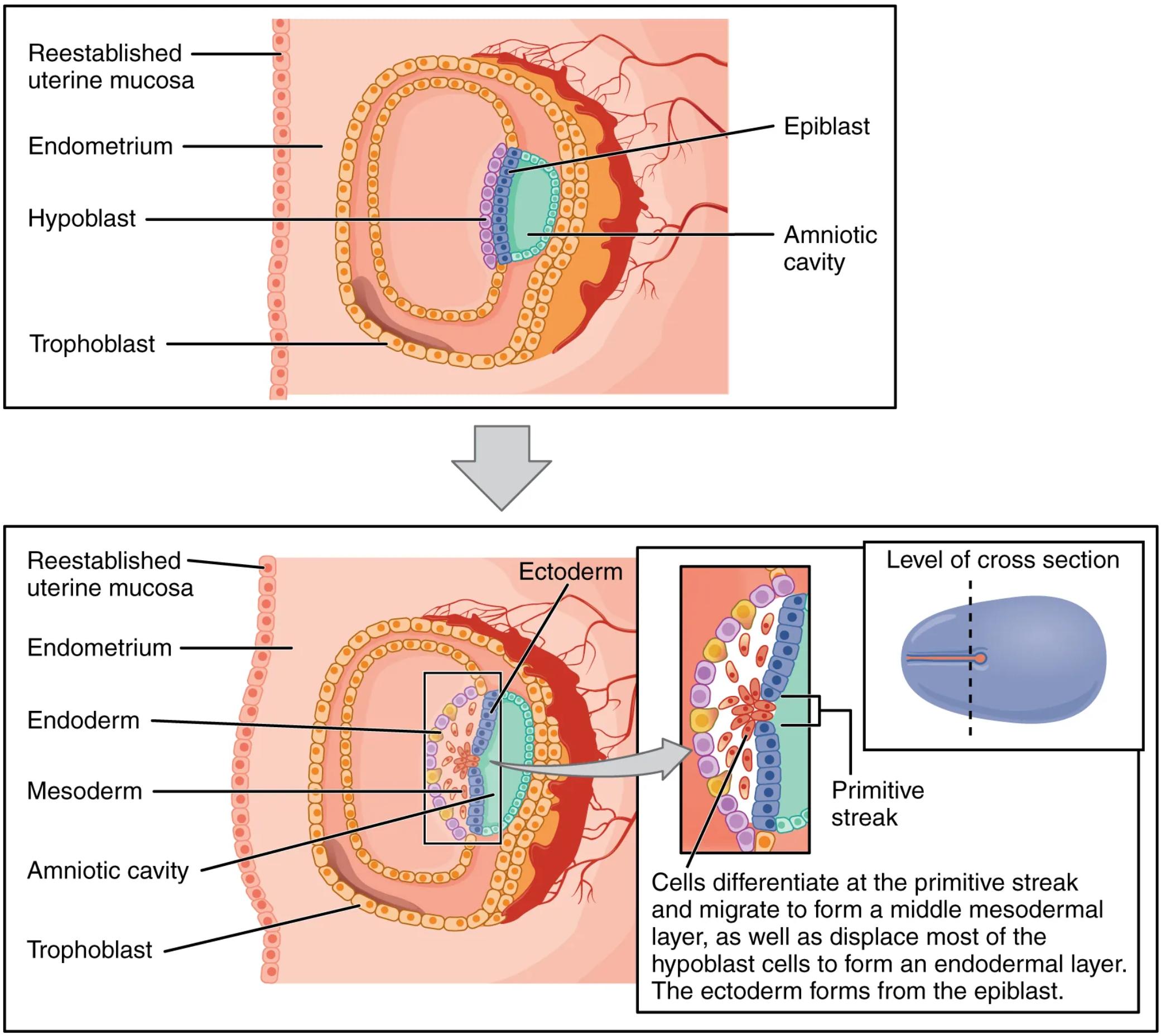
Reestablished uterine mucosa: The innermost lining of the uterus undergoes significant changes throughout the menstrual cycle. Following menstruation, the uterine mucosa rebuilds and becomes receptive to embryo implantation, providing a nutrient-rich environment for the developing embryo.
Endometrium: This is the inner lining of the uterus, which plays a vital role in the menstrual cycle and pregnancy. The endometrium thickens and becomes highly vascularized in preparation for implantation, offering crucial support and sustenance to the conceptus.
Epiblast: A layer of columnar cells within the bilaminar embryonic disc, the epiblast gives rise to the three primary germ layers during gastrulation. It is the source of all embryonic tissues and ultimately forms the embryo proper.
Amniotic cavity: A fluid-filled sac that surrounds and protects the developing embryo. This cavity provides a stable and cushioned environment, allowing for proper growth and movement, and helps to regulate temperature.
Hypoblast: A layer of cuboidal cells located beneath the epiblast, the hypoblast contributes to the formation of the extraembryonic membranes, particularly the yolk sac. While not directly forming embryonic tissues, it plays an important role in early embryonic nutrition and signaling.
Trophoblast: The outer layer of cells of the blastocyst, the trophoblast is essential for implantation and the formation of the placenta. It establishes the critical interface between the maternal uterus and the developing embryo, facilitating nutrient and waste exchange.
Ectoderm: The outermost of the three primary germ layers, the ectoderm differentiates to form the epidermis of the skin, the nervous system (brain and spinal cord), and sensory organs. It is responsible for the development of structures that mediate interaction with the external environment.
Endoderm: The innermost of the three primary germ layers, the endoderm gives rise to the lining of the digestive and respiratory systems, as well as glands such as the liver, pancreas, and thyroid. It forms the internal organs and their associated structures.
Mesoderm: The middle of the three primary germ layers, the mesoderm develops into a wide array of tissues including muscles, bones, cartilage, connective tissue, the circulatory system, and the urogenital system. It is responsible for forming the structural and functional components of the body.
Primitive streak: A transient, linear thickening of epiblast cells that appears on the dorsal surface of the embryonic disc. The primitive streak is a key anatomical landmark during gastrulation, serving as the site where epiblast cells ingress to form the mesoderm and endoderm.
Introduction to Early Embryonic Development
The journey of human development begins with fertilization, followed by a series of rapid cell divisions that lead to the formation of a blastocyst. This blastocyst then implants into the prepared uterine wall, initiating a complex interplay between maternal and embryonic tissues. At this stage, the inner cell mass of the blastocyst differentiates into two distinct layers: the epiblast and the hypoblast, forming what is known as the bilaminar embryonic disc. Surrounding these layers, the trophoblast establishes the crucial connection with the maternal endometrium.
Following implantation, the bilaminar disc undergoes a profound reorganization during a process called gastrulation. This is a pivotal event where the two-layered embryonic disc transforms into a three-layered structure, laying down the fundamental body plan of the embryo. Gastrulation is initiated by the formation of the primitive streak on the dorsal surface of the epiblast. Cells from the epiblast migrate inwards through this streak, undergoing a process of ingression.
During this migration, the first cells to ingress displace the hypoblast, forming the definitive endoderm, which will give rise to the lining of internal organs. Subsequent cells that ingress through the primitive streak spread out between the newly formed endoderm and the remaining epiblast, establishing the mesoderm. This middle layer is critical for forming a vast array of tissues, including muscles, bones, and the circulatory system. The cells remaining in the epiblast after these migrations will then form the ectoderm, which is destined to develop into the nervous system and the outer coverings of the body. This intricate dance of cell movement and differentiation is a hallmark of early embryogenesis, dictating the ultimate fate of every cell in the developing embryo.
The Genesis of the Germ Layers: A Foundation for Organogenesis
The establishment of the three primary germ layers—ectoderm, mesoderm, and endoderm—during gastrulation is a cornerstone of embryonic development. Each layer is pre-programmed to differentiate into specific tissues and organs, a process known as organogenesis. The ectoderm, for instance, not only forms the protective outer skin but also gives rise to the entire central and peripheral nervous system, the sensory organs, and the pituitary gland. This highlights its role in mediating interaction with the external environment and coordinating bodily functions.
The mesoderm, often considered the most diverse of the three, is responsible for the formation of the musculoskeletal system, including all muscles, bones, and cartilage. Furthermore, it generates the circulatory system, complete with the heart, blood vessels, and blood cells, as well as the urogenital system, encompassing the kidneys and reproductive organs. This broad range of derivatives underscores the mesoderm’s importance in providing structural support, movement, and critical physiological systems. Finally, the endoderm forms the lining of the gastrointestinal and respiratory tracts, along with associated glands like the liver, pancreas, and thyroid. These internal linings and glands are vital for nutrient absorption, gas exchange, and hormonal regulation, emphasizing the endoderm’s role in maintaining internal homeostasis.
Understanding the precise sequence of events during gastrulation and the subsequent differentiation of these germ layers is fundamental to embryology. Errors during these early stages can have profound implications for development, leading to various congenital anomalies. Therefore, detailed study of these processes provides invaluable insights into human health and disease. The coordinated molecular and cellular mechanisms that govern germ layer formation represent a remarkable feat of biological organization, transforming a simple bilaminar disc into the foundational blueprint of a complex human being.

