Dracunculiasis, caused by the parasitic nematode Dracunculus medinensis, is a neglected tropical disease that has plagued humanity for centuries, historically referred to as the “fiery serpent.” The accompanying image vividly depicts the traditional and primary clinical method of extraction, where an adult female worm is slowly wound around a small stick to remove it from a painful cutaneous lesion. Understanding this ancient extraction technique and the lifecycle of the parasite is crucial for identifying the disease and appreciating the global eradication efforts currently underway.
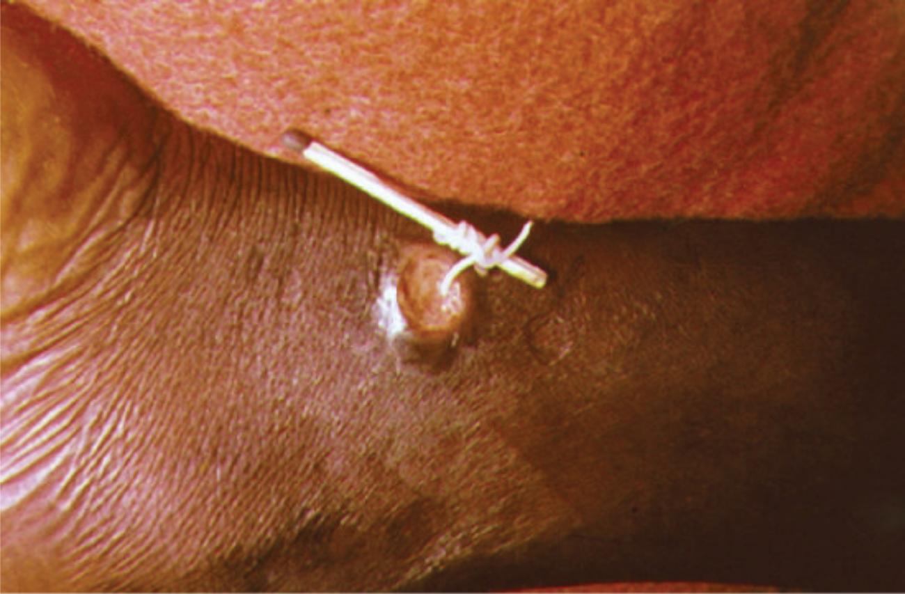
Adult Guinea Worm: This white, filament-like structure is the adult female nematode, which can measure between 60 to 100 centimeters in length despite being only 1 to 2 millimeters wide. It migrates through the subcutaneous tissues to the skin surface specifically to discharge its larvae into water sources, perpetuating its lifecycle.
Matchstick: The small stick serves as a mechanical spool to maintain tension and gradually wind the emerging worm out of the body day by day. This mechanical aid is essential because pulling too hard can cause the worm to snap, leading to the retraction of the remaining segment into the tissue and causing severe inflammation or abscesses.
Cutaneous Lesion: The circular ulcer visible on the skin is the exit site created by the worm’s toxic secretions, which induce a blister that eventually ruptures. This lesion initially causes an intense burning sensation, instinctively prompting the host to submerge the affected limb in water to relieve the pain.
Introduction to the Guinea Worm and Its Impact
The image captures a critical moment in the management of Dracunculiasis, a crippling parasitic infection contracted by drinking stagnant water contaminated with parasite-infected water fleas (copepods). Once ingested, the larvae migrate from the digestive tract into the body cavity, where they mature and mate. The male worms die, but the female worms grow over the course of a year, eventually migrating to the skin to release millions of larvae.
The emergence of the worm is an incredibly painful process. The female worm secretes acid-like substances to create a blister on the skin, usually on the lower limbs. When the person seeks relief by immersing the burning blister in water, the blister bursts, and the worm releases a milky white cloud of larvae into the water, restarting the transmission cycle. The physical extraction shown in the image is currently the only way to remove the parasite, as no drug or vaccine exists to prevent or treat the infection once the worm has matured.
The impact of this disease goes beyond physical pain; it has significant socioeconomic consequences. The emergence of the worm often coincides with the planting or harvesting season in agricultural communities. Because the extraction process can take weeks and renders the patient unable to walk or work, it can lead to food insecurity and economic hardship for affected families.
Key characteristics of the infection include:
- Transmission: Ingestion of water containing infected copepods.
- Incubation Period: Approximately 10 to 14 months before symptoms appear.
- Vector: Cyclops species (microscopic water fleas).
- Primary Symptom: Painful blister and the visible emergence of a white worm.
Pathogenesis and Lifecycle of Dracunculus Medinensis
The lifecycle of Dracunculus medinensis is unique among parasites affecting humans. It begins when a person drinks from a pond or open well containing copepods that carry Guinea worm larvae. Gastric acid in the human stomach digests the copepods, but the larvae survive and penetrate the wall of the intestine. Over the next 10 to 14 months, the female worm grows to a substantial length within the connective tissues, often without causing specific symptoms. This long asymptomatic incubation period makes early diagnosis difficult.
As the female worm becomes gravid (filled with embryos), she migrates toward the skin surface, typically favoring the legs and feet. The worm provokes a localized immune response, resulting in a painful papule that evolves into a blister. The rupture of this blister exposes the anterior end of the worm. This process is evolutionarily adapted to ensure the parasite reaches water, as the burning pain drives the host to cool the wound, thereby allowing the worm to eject its larvae into the water source.
Clinical Management and Extraction Techniques
There is no pharmacological cure for Dracunculiasis once the adult worm has established itself in the subcutaneous tissue. Anthelmintic drugs usually effective against other worms are ineffective here. Instead, management relies on the careful physical removal of the parasite, as demonstrated in the image. The standard procedure involves winding the worm a few centimeters each day around a small stick or piece of gauze. This process is slow and requires patience; extracting a single worm can take anywhere from a few days to several weeks.
Medical care during this period focuses on preventing secondary bacterial infections. The open wound is a gateway for bacteria, which can lead to cellulitis, abscesses, or even tetanus. Therefore, the lesion must be cleaned and dressed regularly. Pain management is also a priority, as the inflammation caused by the worm’s movement and the open ulcer can be debilitating. If the worm breaks during extraction, the dead portion remaining inside the body degrades, causing a severe inflammatory reaction that can lead to permanent scarring or joint deformities (arthritis) if the worm is located near a joint.
The Success of Global Eradication Efforts
While the image depicts a painful reality, it also represents a disease on the brink of eradication. Through the use of simple filtration devices (like pipe filters and nylon cloth) to remove copepods from drinking water, active surveillance, and vector control, the number of global cases has dropped from millions in the 1980s to only a handful in recent years. This success story highlights the power of public health interventions that focus on behavioral change and water safety rather than complex medical treatments.
Conclusion
The extraction of Dracunculus medinensis using a simple stick remains the definitive image of Dracunculiasis. It represents a collision between human physiology and a highly specialized parasite that exploits the human need for water to survive. For healthcare professionals, recognizing this distinct presentation is vital, particularly in endemic regions. The continued vigilance in detecting and managing these cases is the final hurdle in making Guinea worm disease the first parasitic disease to be eradicated from the human population.

