The foot’s superficial musculature on the dorsal side plays a vital role in facilitating movement and maintaining stability, particularly along its lateral aspect. This article examines the dorsal superficial muscles of the right foot, presented in a lateral view, to provide a detailed look at their anatomical structure and functional contributions. These muscles, primarily responsible for extending the toes, enhance the foot’s ability to adapt to various surfaces and support dynamic activities. By exploring the labeled diagram, readers can gain a comprehensive understanding of these muscles’ significance in foot function and their relevance in clinical settings.
Introduction to the Dorsal Superficial Muscles
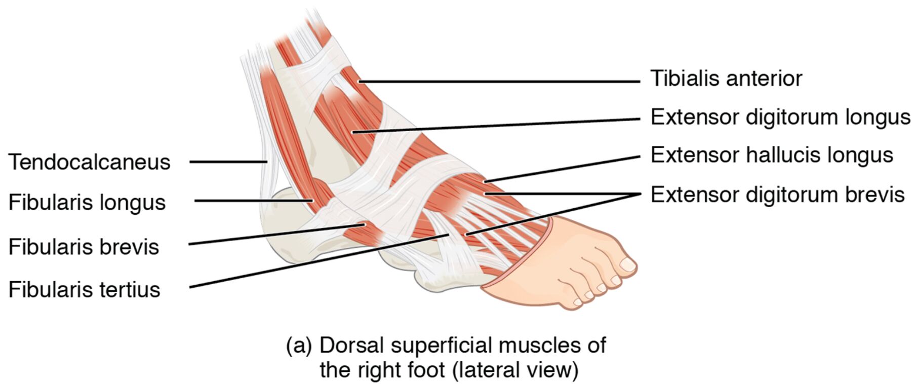
The dorsal superficial muscles of the right foot form the outer layer on the top of the foot. Their lateral view highlights their role in toe movement and stability. This section details the labeled structures that define their anatomy and function.
- Extensor digitorum brevis: Positioned on the lateral dorsal side, this muscle extends the toes. It assists in lifting the toes during the swing phase of walking.
- Extensor hallucis brevis: Located near the big toe on the dorsal surface, it extends the big toe. It supports precise toe movements and foot balance.
- Extensor digitorum longus (tendon): Found laterally as a tendon, it extends the toes and dorsiflexes the foot. It transmits force from the lower leg to the foot.
- Extensor hallucis longus (tendon): Positioned as a tendon near the big toe, it extends the big toe and dorsiflexes the foot. It enhances toe lifting and stability.
- Superior extensor retinaculum: A band on the lateral dorsal ankle, it holds extensor tendons in place. It ensures smooth tendon movement during dorsiflexion.
- Inferior extensor retinaculum: Another band on the lateral dorsal ankle, it stabilizes extensor tendons. It prevents tendon displacement during foot motion.
The dorsal superficial muscles of the right foot‘s lateral placement optimizes toe function. Their labeled view provides a clear perspective on their structural and operational roles.
Functional Roles of the Dorsal Superficial Muscles
The dorsal superficial muscles of the right foot are essential for toe extension and foot movement. Their actions support mobility and coordination. This section outlines their specific functional contributions.
- The extensor digitorum brevis extends the toes, facilitating lifting during gait. It enhances foot clearance and balance on uneven surfaces.
- The extensor hallucis brevis extends the big toe, improving toe-off precision. It contributes to stable foot positioning during movement.
- The extensor digitorum longus (tendon) extends the toes and dorsiflexes the foot. This action aids in smooth transitions between steps.
- The extensor hallucis longus (tendon) extends the big toe and dorsiflexes the foot. It supports toe elevation and overall foot flexibility.
- The superior extensor retinaculum and inferior extensor retinaculum stabilize the extensor tendons. They ensure efficient force transmission during extension.
The dorsal superficial muscles of the right foot‘s coordinated efforts enhance foot performance. Their lateral focus supports dynamic and stable movement.
Clinical Significance and Practical Applications
The dorsal superficial muscles of the right foot are often evaluated in clinical assessments of foot health. Their condition directly impacts mobility and stability. This section explores their clinical relevance.
- Strain in the extensor digitorum brevis can lead to toe extension pain. Stretching and strengthening exercises help restore function and flexibility.
- Weakness in the extensor hallucis brevis may impair big toe movement, affecting push-off. Targeted therapy improves toe strength and stability.
- Injury to the extensor digitorum longus (tendon) can cause dorsiflexion issues. Rehabilitation focuses on tendon repair and foot lifting ability.
- Overuse of the extensor hallucis longus (tendon) may result in tendonitis, limiting toe extension. Rest and conditioning prevent further damage.
- Understanding their anatomy aids in diagnosing conditions like anterior ankle impingement. This knowledge guides effective treatment and preventive measures.
This insight is valuable for professionals addressing foot concerns. The dorsal superficial muscles of the right foot‘s roles underscore the need for precise therapeutic interventions.
Conclusion
The dorsal superficial muscles of the right foot, as depicted in the lateral view, illustrate the foot’s intricate muscular design on its dorsal surface. This article has explored their anatomical structure, diverse functional roles, and clinical significance, providing a thorough understanding of their importance. From the extensor digitorum brevis extending the toes to the extensor hallucis longus (tendon) supporting dorsiflexion, each muscle contributes uniquely to foot mobility and stability. Continued study of these muscles will enhance therapeutic strategies and deepen appreciation for the complex mechanics of foot movement.

