The larynx, a key component of the airway system, extends from the laryngopharynx to the trachea, playing an indispensable role in breathing, voice production, and swallowing. Positioned beneath the hyoid bone, this cartilaginous structure houses the vocal cords and protects the trachea through its robust yet flexible design. A right lateral view of the larynx provides a detailed perspective on its anatomy, offering insights into its functional and structural intricacies.
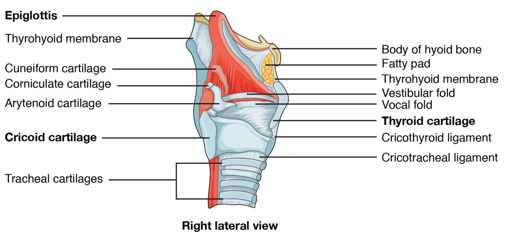
Key Anatomical Labels in the Diagram
This section explores each labeled component, providing clarity on their roles and positions within the laryngeal framework.
Epiglottis: The epiglottis is a leaf-like cartilage that seals the larynx during swallowing, preventing food from entering the airway. It opens during respiration to allow air to flow into the trachea.
Thyrohyoid membrane: The thyrohyoid membrane connects the thyroid cartilage to the hyoid bone, supporting laryngeal elevation during swallowing. It contains the internal laryngeal nerve, which provides sensory innervation.
Cuneiform cartilage: The cuneiform cartilage is a small, rod-shaped structure within the aryepiglottic fold, supporting the epiglottis’s shape. It contributes to the stability of the laryngeal inlet.
Corniculate cartilage: The corniculate cartilage sits atop the arytenoid cartilage, aiding in closing the laryngeal inlet during swallowing. It enhances the epiglottis’s protective function.
Arytenoid cartilage: The arytenoid cartilage is a paired structure on the cricoid cartilage, controlling vocal cord movement through muscle action. It rotates to adjust the glottis for breathing or phonation.
Cricoid cartilage: The cricoid cartilage forms a complete ring below the thyroid cartilage, serving as the larynx’s foundation. It supports the airway and anchors the esophagus posteriorly.
Tracheal cartilages: The tracheal cartilages are C-shaped rings extending from the cricoid cartilage, maintaining tracheal openness. They prevent collapse during respiration and guide air to the lungs.
Body of hyoid bone: The body of the hyoid bone is a U-shaped structure above the larynx, anchoring tongue and laryngeal muscles. It remains unattached to other bones, facilitating speech and swallowing.
Fatty pad: The fatty pad within the thyrohyoid membrane cushions the larynx against external pressure. It also contributes to the smooth contour of the neck.
Thyrohyoid membrane: The thyrohyoid membrane stretches between the thyroid cartilage and hyoid bone, aiding laryngeal movement. It houses nerves and vessels critical for sensory and motor functions.
Vestibular fold: The vestibular fold, or false vocal cord, lies above the true vocal cord, protecting the airway during swallowing. It plays a minor role in sound production but aids in closure.
Vocal fold: The vocal fold, or true vocal cord, vibrates to produce sound as air passes, controlled by the recurrent laryngeal nerve. It is essential for speech and the cough reflex.
Thyroid cartilage: The thyroid cartilage forms the laryngeal prominence or Adam’s apple, encasing the vocal cords. It connects to the hyoid bone, providing structural support.
Cricothyroid ligament: The cricothyroid ligament links the thyroid and cricoid cartilages, allowing vocal pitch adjustment. It serves as an emergency airway access point.
Cricotracheal ligament: The cricotracheal ligament connects the cricoid cartilage to the trachea, ensuring a flexible transition. It maintains airway continuity and stability.
Cartilaginous Components and Their Roles
The larynx’s cartilage provides a sturdy yet adaptable framework. This structure supports its critical functions effectively.
- The thyroid cartilage protects the vocal cords, with its prominence varying by gender.
- Cricoid cartilage offers a stable base, unique for its complete ring.
- Arytenoid cartilage controls vocal fold movement, enabling phonation.
- Epiglottis and cuneiform cartilages safeguard the airway during swallowing.
- Tracheal cartilages ensure an open passage to the lungs.
Ligaments and Membranes in Laryngeal Function
Ligaments and membranes enhance the larynx’s mobility and stability. They connect key structures, ensuring coordinated action.
- The thyrohyoid membrane elevates the larynx, aiding swallowing.
- Cricothyroid ligament adjusts vocal pitch through muscle tension.
- Cricotracheal ligament maintains a smooth tracheal-laryngeal junction.
- The fatty pad cushions the larynx, protecting against trauma.
- These elements adapt to pressure changes during respiration.
Functional Significance in Respiration and Phonation
The larynx serves as a multifunctional organ, vital for breathing and voice. Its anatomy supports diverse physiological needs.
- The epiglottis prevents aspiration, directing food away from the trachea.
- Vocal folds vibrate to produce sound, modulated by arytenoid movement.
- The laryngeal prominence shields the delicate vocal apparatus.
- Tracheal cartilages maintain airway patency under varying conditions.
- The hyoid bone anchors muscles, supporting speech articulation.
Clinical Implications and Anatomical Variations
Understanding laryngeal anatomy aids in diagnosing and managing related conditions. Variations can influence clinical outcomes.
- Epiglottitis can obstruct the airway, requiring immediate intervention.
- Vocal fold paralysis, due to nerve damage, affects phonation.
- Thyroid cartilage fractures from trauma may impair voice.
- Cricothyroid ligament access is critical in emergency tracheostomies.
- Imaging helps assess structural deviations for surgical planning.
The larynx’s intricate design, from the epiglottis to the tracheal cartilages, highlights its role in respiration, phonation, and airway protection. By examining its anatomy through the right lateral view, one gains a deeper appreciation for its contributions to human physiology, showcasing the precision of this essential structure.

