The intricate process of early human development is a marvel of biological precision, transforming a single-celled zygote into a complex multicellular embryo. This image provides a crucial glimpse into the formation of the embryonic disc, a foundational structure from which the entire organism will arise. Understanding these initial stages is vital for comprehending the basis of human anatomy and the potential origins of developmental anomalies.
Key Structures of the Embryonic Disc and Surrounding Tissues
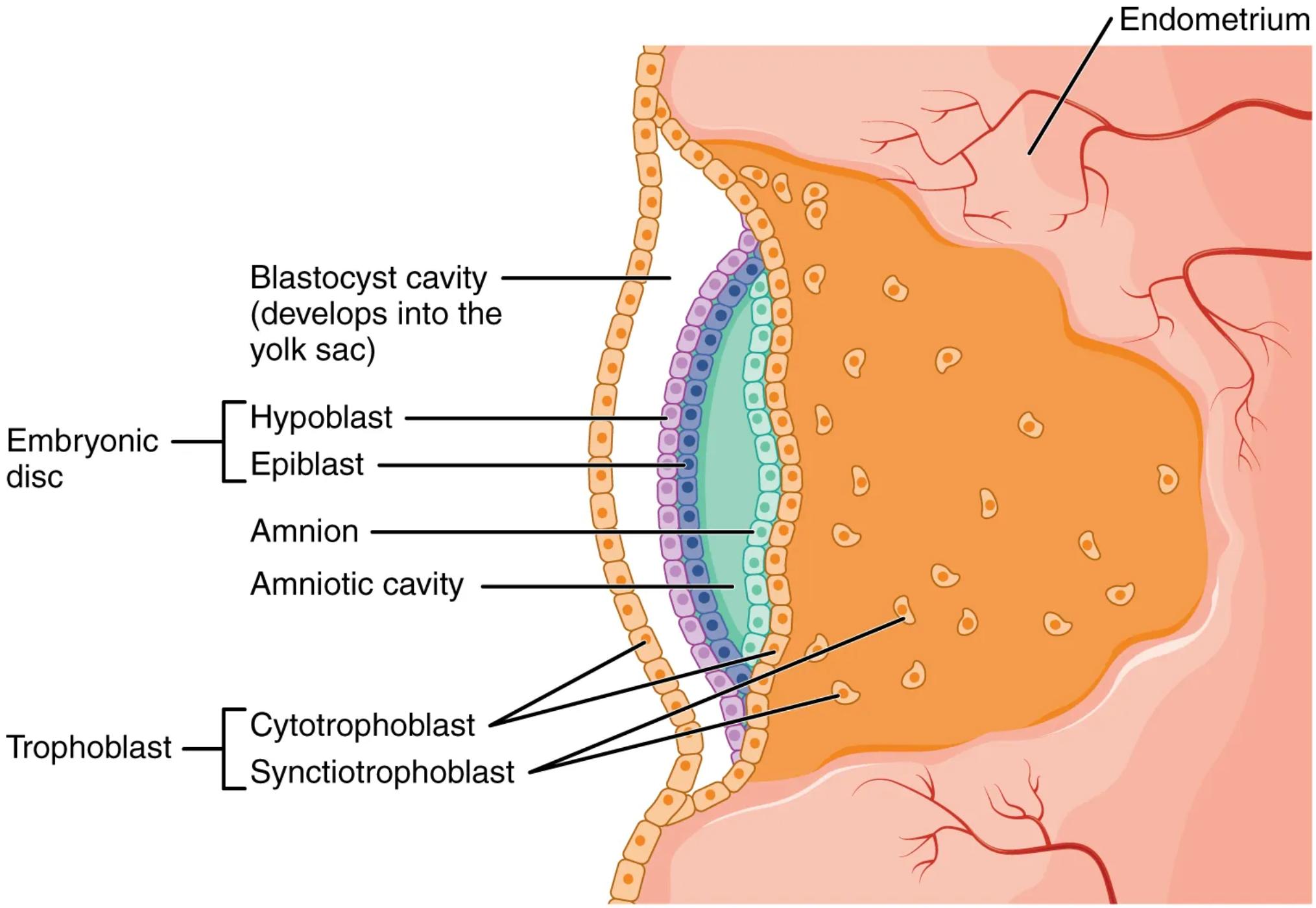
Here’s a detailed explanation of the labeled components shown in the image:
Endometrium: This refers to the inner lining of the uterus. It undergoes cyclical changes in preparation for potential embryo implantation and provides a nutrient-rich environment for the developing blastocyst.
Blastocyst cavity (develops into the yolk sac): Initially a fluid-filled space within the blastocyst, this cavity later contributes to the formation of the yolk sac. The yolk sac plays a critical role in early embryonic nutrition and the development of primordial germ cells.
Hypoblast: This is one of the two primary cell layers forming the embryonic disc, located ventral to the epiblast. The hypoblast contributes to the formation of the yolk sac and is crucial for signaling during early development.
Epiblast: The second primary cell layer of the embryonic disc, situated dorsal to the hypoblast. The epiblast is multipotent and gives rise to all three germ layers (ectoderm, mesoderm, and endoderm) of the embryo proper.
Amnion: This is a membrane that forms the wall of the amniotic cavity. It encloses the developing embryo and amniotic fluid, providing protection and allowing for fetal movement.
Amniotic cavity: A fluid-filled sac that surrounds and cushions the developing embryo. The amniotic fluid within it helps regulate temperature, prevents adhesion of the embryo to the amnion, and aids in musculoskeletal development.
Cytotrophoblast: The inner layer of the trophoblast, consisting of distinct cells that divide and contribute to the formation of the syncytiotrophoblast. It plays a vital role in anchoring the blastocyst to the endometrium.
Syncytiotrophoblast: The outer, multinucleated layer of the trophoblast that invades the endometrium of the uterus. It is responsible for producing hormones like human chorionic gonadotropin (hCG) and facilitating nutrient exchange between the mother and the developing embryo.
The Genesis of Life: Understanding the Embryonic Disc
The image beautifully illustrates a pivotal moment in human embryogenesis, approximately during the second week post-fertilization, where the blastocyst has successfully implanted into the endometrium of the uterus. At this stage, the inner cell mass of the blastocyst differentiates into two distinct layers, the epiblast and the hypoblast, which together constitute the bilaminar embryonic disc. This disc is the precursor to the entire embryo and its associated membranes.
On either side of this embryonic disc, two crucial cavities begin to form. Dorsal to the epiblast, the amniotic cavity emerges, destined to envelop the growing embryo in protective amniotic fluid. Ventral to the hypoblast, the blastocyst cavity expands and reorganizes to become the primary yolk sac. While the human yolk sac does not contain significant nutritional reserves like in other species, it serves as an important site for early blood cell formation and germ cell development before these functions are taken over by other organs.
The interaction between the invading trophoblast and the maternal endometrium is fundamental for successful implantation and the establishment of pregnancy. The syncytiotrophoblast, with its aggressive invasive properties, penetrates the maternal tissues, creating a secure attachment for the developing embryo and initiating the formation of the placenta. Concurrently, the cytotrophoblast provides a steady supply of new cells to the expanding syncytiotrophoblast and forms a protective layer around the nascent embryo. This coordinated cellular activity ensures the embryo’s continued growth and development within the maternal environment.
Understanding these early stages, particularly the precise formation and function of the embryonic disc and its surrounding structures, is paramount in developmental biology. Deviations during this delicate period can lead to various congenital anomalies, highlighting the importance of normal cellular differentiation, migration, and tissue organization. This intricate dance of cells and tissues lays the groundwork for all subsequent embryonic and fetal development, ultimately leading to the formation of a complete human being.
Conclusion
The image presented offers an invaluable window into the complex and highly orchestrated events of early human development. From the initial differentiation of the epiblast and hypoblast to the formation of the amniotic cavity and yolk sac, each component plays a specific and vital role in establishing the blueprint for the entire organism. The dynamic interplay between the trophoblast layers and the endometrium underscores the critical maternal-fetal interface that supports the embryo’s survival and growth. A thorough grasp of these fundamental processes is essential for anyone delving into the intricacies of embryology and reproductive health.

