The lower leg’s deep musculature plays a crucial role in supporting intricate movements and maintaining foot stability, often hidden beneath the superficial layers. This article explores the deep muscles of the right lower leg, presented in a posterior view, to provide a detailed examination of their anatomical structure and functional importance. These muscles, primarily located in the posterior compartment, are responsible for plantar flexion, with additional contributions to inversion, eversion, and rotation of the foot through lateral and medial actions. By analyzing the labeled diagram, readers can gain a comprehensive insight into these muscles’ roles in leg function and their significance in clinical contexts.
Introduction to the Deep Muscles of the Lower Leg
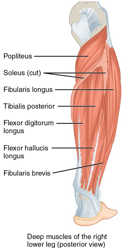
The deep muscles of the right lower leg lie beneath the surface, offering essential support in the posterior compartment. Their posterior view reveals a complex network critical for foot and leg dynamics. This section details the labeled structures that define their anatomy and function.
- Popliteus: Positioned deep in the posterior compartment, this muscle flexes and internally rotates the knee. It unlocks the knee joint during movement initiation.
- Tibialis posterior: Located medially in the deep posterior layer, it inverts and plantar flexes the foot. It maintains the medial arch and supports foot stability.
- Flexor digitorum longus: Found deep posteriorly, this muscle flexes the toes and plantar flexes the foot. It aids in gripping the ground during push-off.
- Flexor hallucis longus: Positioned deep in the posterior compartment, it flexes the big toe and plantar flexes the foot. It enhances toe stability during locomotion.
- Fibularis longus: Located laterally in the deep posterior view, it everts and plantar flexes the foot. It provides lateral ankle support and stability.
- Calcaneal (Achilles) tendon: Connects deep calf muscles to the heel, it transmits force for plantar flexion. It is vital for efficient movement and propulsion.
- Calcaneus (heel): The heel bone serves as an insertion point for the Achilles tendon. It anchors the deep posterior muscles for effective action.
The deep muscles of the right lower leg‘s posterior placement ensures robust support. Their labeled anatomy offers a detailed perspective on their structural and functional roles.
Functional Roles of the Deep Muscles
The deep muscles of the right lower leg are essential for precise foot and leg movements. Their actions in the posterior compartment enhance stability and mobility. This section outlines their specific functional contributions.
- The tibialis posterior inverts and plantar flexes the foot, maintaining the medial arch. It supports foot alignment during weight-bearing activities.
- The flexor digitorum longus flexes the toes and plantar flexes the foot. This action aids in gripping the ground and enhancing push-off strength.
- The flexor hallucis longus flexes the big toe and plantar flexes the foot. It provides stability to the toe during the toe-off phase of gait.
- The popliteus flexes and internally rotates the knee, unlocking the joint. This movement facilitates smooth transitions from standing to bending.
- The fibularis longus everts and plantar flexes the foot, supporting lateral stability. It assists in balancing on uneven surfaces.
- The calcaneal tendon transmits force from deep muscles to the heel. This ensures powerful plantar flexion during dynamic movements.
The deep muscles of the right lower leg‘s coordinated efforts optimize leg performance. Their deep location provides critical support for complex motions.
Clinical Significance and Practical Applications
The deep muscles of the right lower leg are often evaluated in clinical assessments of leg and foot health. Their condition directly influences mobility and daily function. This section explores their clinical relevance.
- Strain in the tibialis posterior can lead to flat feet or posterior tibial tendon dysfunction. Strengthening exercises help restore arch support and stability.
- Injury to the flexor hallucis longus may impair big toe flexion, affecting push-off. Physical therapy targets this muscle to regain toe function.
- Weakness in the flexor digitorum longus can reduce toe grip, impacting balance. Targeted training improves toe flexion and ground contact.
- Damage to the popliteus may cause knee instability or pain. Rehabilitation focuses on restoring knee flexion and rotation.
- Understanding their anatomy aids in diagnosing conditions like deep posterior compartment syndrome. This knowledge guides effective treatment and prevention strategies.
This insight is valuable for professionals addressing leg issues. The deep muscles of the right lower leg‘s roles underscore the need for precise therapeutic interventions.
Conclusion
The deep muscles of the right lower leg, as illustrated in the posterior view, reveal the intricate support system beneath the surface of the lower limb. This article has explored their anatomical structure, diverse functional roles, and clinical significance, providing a thorough understanding of their importance. From the tibialis posterior maintaining foot arches to the popliteus unlocking the knee, each muscle contributes uniquely to leg stability and movement. Continued study of these muscles will enhance therapeutic approaches and deepen appreciation for the complex mechanics of the lower leg.

