The deep musculature of the foot’s sole forms a critical foundation for supporting weight and enabling intricate movements, lying beneath the surface layers. This article delves into the deep muscles of the left sole, presented in a plantar view, to provide a detailed examination of their anatomical structure and functional roles within the third and deepest layer of the plantar region. These muscles, primarily responsible for flexing the toes and stabilizing the foot’s arches, play a vital role in counterbalancing body weight and facilitating locomotion. By analyzing the labeled diagram, readers can gain a comprehensive understanding of these muscles’ significance in foot function and their relevance in clinical settings.
Introduction to the Deep Muscles of the Sole
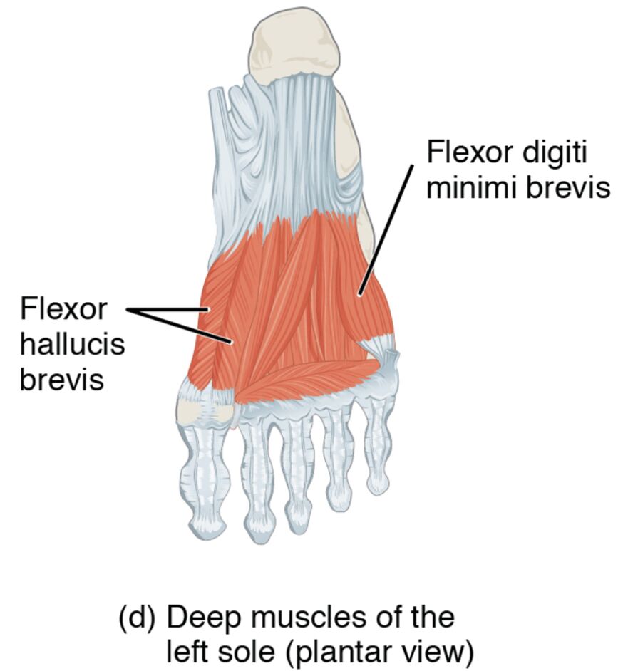
The deep muscles of the left sole reside in the innermost layer of the plantar surface. Their plantar view reveals their essential contribution to foot strength and movement. This section details the labeled structures that define their anatomy and function.
- Flexor hallucis brevis: Positioned near the big toe, this muscle flexes the proximal phalanx of the big toe. It supports propulsion and maintains toe stability during push-off.
- Adductor hallucis (oblique head): Located medially, it adducts and flexes the big toe. It stabilizes the toe and reinforces the transverse arch.
- Adductor hallucis (transverse head): Found across the metatarsal heads, it adducts the big toe and supports the transverse arch. It aids in maintaining foot alignment under load.
- Flexor digiti minimi brevis: Positioned near the little toe, it flexes the proximal phalanx of the little toe. It enhances lateral toe movement and stability.
- Plantar interossei: Located between the metatarsals, these muscles adduct the toes toward the third toe. They assist in fine-tuning toe positioning and balance.
- Dorsal interossei: Positioned between the metatarsals, these muscles abduct the toes away from the third toe. They contribute to toe spreading and lateral stability.
The deep muscles of the left sole‘s deep placement ensures robust support. Their labeled anatomy provides a detailed perspective on their structural and functional roles.
Functional Roles of the Deep Muscles
The deep muscles of the left sole are crucial for precise toe movements and foot support. Their positions in the deepest plantar layer enhance stability and flexibility. This section outlines their specific functional contributions.
- The flexor hallucis brevis flexes the big toe, powering the push-off phase. It also supports the longitudinal arch during weight-bearing.
- The adductor hallucis (oblique head) and adductor hallucis (transverse head) adduct the big toe, reinforcing arch stability. They enhance propulsion and load distribution.
- The flexor digiti minimi brevis flexes the little toe, improving lateral grip. It aids in maintaining balance on uneven surfaces.
- The plantar interossei adduct the toes, fine-tuning their alignment. This action supports precise movements and weight distribution.
- The dorsal interossei abduct the toes, promoting toe spreading. They enhance lateral stability and foot adaptability.
The deep muscles of the left sole‘s coordinated efforts optimize foot performance. Their deep location provides critical support for complex motions.
Clinical Significance and Practical Applications
The deep muscles of the left sole are often evaluated in clinical assessments of foot health. Their condition directly impacts mobility and stability. This section explores their clinical relevance.
- Weakness in the flexor hallucis brevis can lead to big toe instability or hallux rigidus. Strengthening exercises help restore toe function and arch support.
- Strain in the adductor hallucis may cause toe pain or bunion formation. Stretching and conditioning alleviate discomfort and improve alignment.
- Injury to the flexor digiti minimi brevis can impair little toe flexion, affecting lateral stability. Targeted therapy restores movement and balance.
- Overuse of the plantar interossei or dorsal interossei may contribute to metatarsalgia. Rest and rehabilitation prevent further strain and enhance comfort.
- Understanding their anatomy aids in diagnosing conditions like Morton’s neuroma. This knowledge guides effective treatment and preventive strategies.
This insight is valuable for professionals addressing foot issues. The deep muscles of the left sole‘s roles underscore the need for precise therapeutic interventions.
Conclusion
The deep muscles of the left sole, as depicted in the plantar view, reveal the foot’s intricate muscular framework that supports toe movement and stability in its deepest layer. This article has explored their anatomical structure, diverse functional roles, and clinical significance, providing a thorough understanding of their importance. From the flexor hallucis brevis enhancing push-off to the dorsal interossei promoting toe spreading, each muscle contributes uniquely to foot function and balance. Continued study of these muscles will enhance therapeutic approaches and deepen appreciation for the complex mechanics of the foot.

