Deep Muscles of the Back and Spinal Muscles: Anatomy and Function
The human back is a complex structure supported by a network of deep muscles that play a critical role in maintaining posture, facilitating movement, and stabilizing the spine. This detailed anatomical image highlights the deep muscles of the back and deep spinal muscles, offering a clear view of their arrangement and function in the neck, shoulders, and vertebral column. Understanding these muscles is essential for grasping how the body supports its upper structure and enables a range of motions, from simple head turns to complex spinal adjustments.
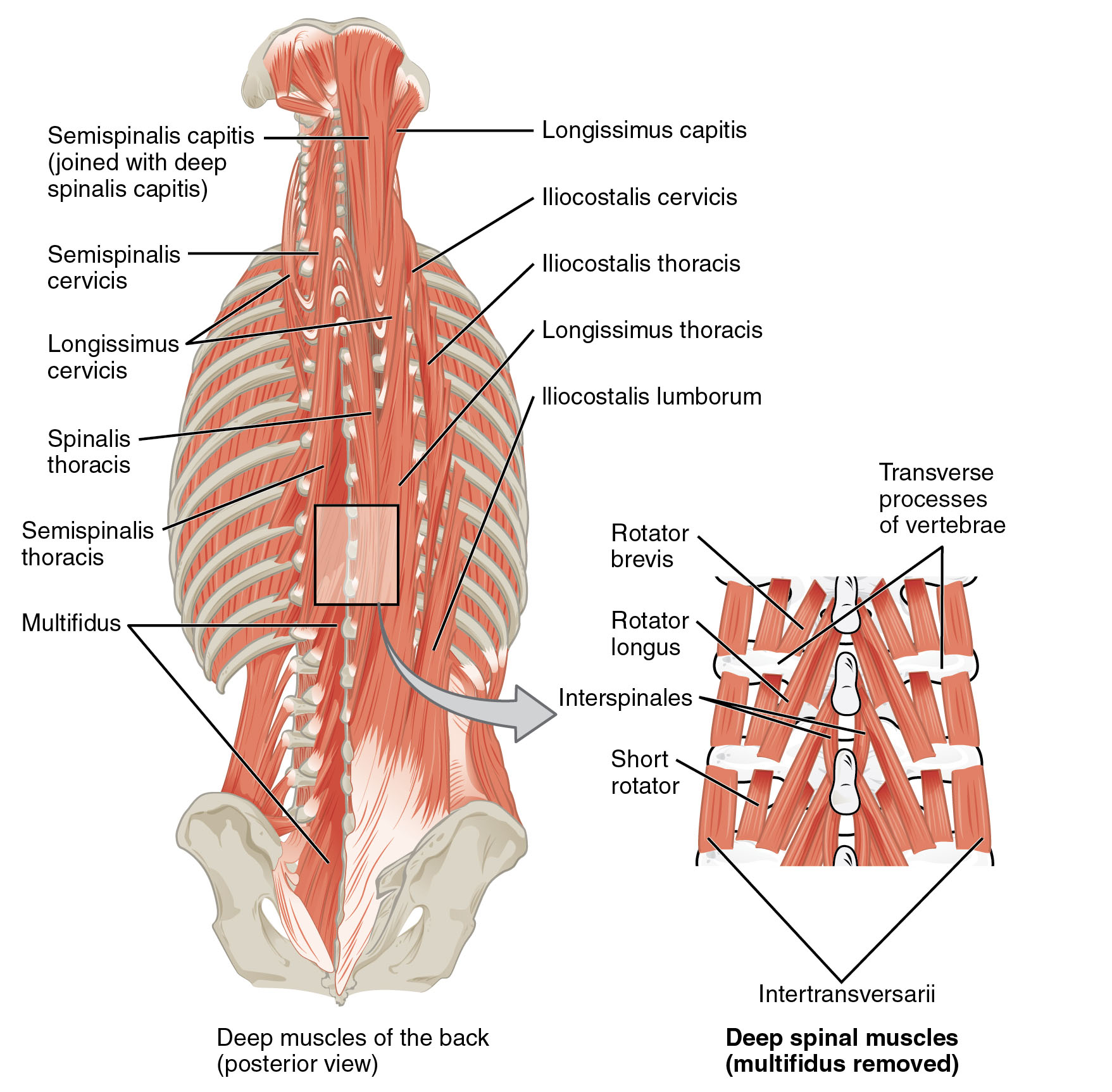
Labels Introduction
- Semispinalis capitis (joined with deep spinalis capitis)
This muscle is located at the upper back of the neck and works to extend and rotate the head. It collaborates with the deep spinalis capitis to provide additional support and stability to the cervical spine. - Semispinalis cervicis
Positioned in the cervical region, this muscle aids in extending and rotating the neck, contributing to smooth head movements. It plays a vital role in maintaining the alignment of the upper spine during various activities. - Longissimus cervicis
Found along the cervical spine, this muscle assists in extending and laterally flexing the neck, ensuring proper posture and movement. It is part of the larger erector spinae group, which supports the vertebral column. - Semispinalis thoracis
Located in the thoracic region, this muscle extends and rotates the upper back, providing stability to the thoracic spine. It helps in coordinating movements involving the rib cage and upper torso. - Longissimus thoracis
This muscle runs along the thoracic spine and aids in extending and laterally flexing the back, supporting upright posture. It is a key component in maintaining the structural integrity of the mid-back. - Iliocostalis cervicis
Positioned in the cervical area, this muscle assists in extending and stabilizing the neck, working in tandem with other spinal muscles. It helps prevent excessive strain during head movements. - Iliocostalis thoracis
Located in the thoracic region, this muscle supports the extension and lateral flexion of the upper back, contributing to respiratory movements. It connects the ribs to the spine, aiding in breathing mechanics. - Iliocostalis lumborum
Found in the lumbar region, this muscle extends and laterally flexes the lower back, providing essential support to the lumbar spine. It plays a crucial role in maintaining lumbar stability during lifting or bending. - Multifidus
This deep muscle spans the entire length of the spine, stabilizing individual vertebrae and facilitating rotation and extension. Its segmented structure allows for precise control of spinal movements. - Rotator brevis
Located in the deep spinal muscles, this muscle rotates the spine and assists in maintaining vertebral alignment. It works closely with other rotators to ensure smooth spinal motion. - Rotator longus
Positioned among the deep spinal muscles, this muscle aids in rotating the spine and provides additional support to the vertebral column. It enhances the spine’s ability to twist and turn effectively. - Interspinales
These small muscles lie between the spinous processes of vertebrae, assisting in extending and stabilizing the spine. They contribute to fine-tuned adjustments of spinal posture. - Short rotator
Found within the deep spinal muscles, this muscle facilitates spinal rotation and helps maintain vertebral integrity. It supports the spine during dynamic movements. - Intertransversarii
Located between the transverse processes of vertebrae, these muscles assist in lateral flexion and stabilization of the spine. They play a subtle yet important role in side-to-side movements. - Transverse processes of vertebrae
These bony projections serve as attachment points for the deep spinal muscles, providing structural support to the spine. They are critical for the leverage and stability of spinal movements.
Anatomical Overview of Deep Back Muscles
The deep muscles of the back form a sophisticated network that underpins the body’s structural integrity. These muscles, including the semispinalis, longissimus, and iliocostalis groups, are part of the erector spinae, which runs parallel to the spine. Their primary function is to extend and stabilize the vertebral column, ensuring the body can maintain an upright posture. Additionally, muscles like the multifidus and intertransversarii provide segmental support, allowing for precise control over individual vertebrae.
The cervical region, where muscles such as the semispinalis capitis and longissimus cervicis reside, is particularly important for head movement and neck stability. These muscles work in unison to allow for rotation, extension, and lateral flexion, which are essential for daily activities like looking over the shoulder. In the thoracic area, the iliocostalis thoracis and semispinalis thoracis not only support the spine but also assist in respiratory mechanics by facilitating rib movement during breathing.
Moving to the lumbar region, the iliocostalis lumborum and multifidus provide critical support to the lower back, a common site of strain and injury. These muscles help absorb shock and maintain lumbar lordosis, the natural inward curve of the lower spine. The deep spinal muscles, including the rotator brevis, rotator longus, and interspinales, further enhance spinal mobility by enabling rotation and fine adjustments, ensuring the spine remains flexible yet stable.
Functional Role in Movement and Posture
The deep muscles of the back are integral to a wide range of movements and postural adjustments. The longissimus thoracis and iliocostalis cervicis, for instance, contribute to lateral flexion, allowing the body to bend sideways, which is vital for activities like stretching or reaching. Extension of the spine, facilitated by the semispinalis thoracis and multifidus, is crucial for standing up straight or arching the back, movements that engage the entire posterior chain.
Rotation, a complex motion, relies heavily on the rotator brevis and short rotator, which work to twist the spine with precision. This capability is essential for turning the torso or looking behind without shifting the lower body. The intertransversarii and interspinales play a supportive role by stabilizing the spine during these dynamic actions, preventing excessive strain on the vertebral joints.
Posture is another area where these muscles shine, with the iliocostalis lumborum and longissimus cervicis maintaining the natural curves of the spine. Poor posture can lead to overcompensation by these muscles, potentially causing fatigue or discomfort. Regular strengthening and stretching exercises targeting these muscle groups can enhance spinal health and prevent chronic issues.
Clinical Relevance and Muscle Health
Maintaining the health of the deep back muscles is crucial for overall musculoskeletal well-being. Strain or injury to the semispinalis capitis or multifidus, for example, can result from poor lifting techniques or prolonged sitting, leading to pain or reduced mobility. Physical therapy often focuses on strengthening these muscles to alleviate lower back pain, a prevalent condition affecting many individuals.
The transverse processes of vertebrae serve as anchor points for these muscles, and any misalignment can affect muscle function, potentially leading to conditions like scoliosis or herniated discs. Regular assessment by healthcare professionals can help identify imbalances early, allowing for targeted interventions. Incorporating exercises that engage the erector spinae and deep spinal muscles can promote long-term spinal health and resilience.
Conclusion
The deep muscles of the back and spine form a remarkable system that supports the body’s structural and functional needs. From the semispinalis cervicis in the neck to the iliocostalis lumborum in the lower back, each muscle contributes to a harmonious balance of movement and stability. Understanding their anatomy and role can guide effective exercise routines and therapeutic approaches, ensuring a healthy and active lifestyle. By appreciating the complexity of these muscles, individuals can take proactive steps to support their spinal health and overall well-being.

