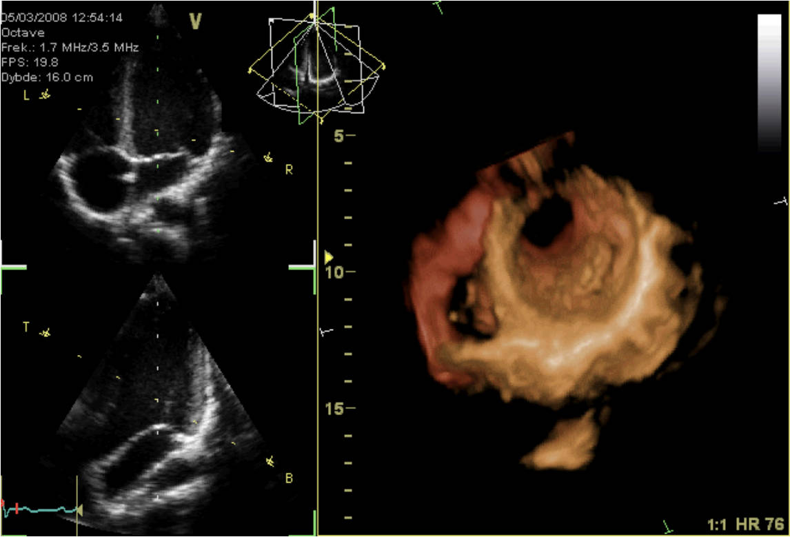
This image presents a sophisticated look at cardiac imaging, combining 2D echocardiogram views with a 3D reconstruction of the heart. Such advanced diagnostic tools are vital for non-invasively assessing heart structure and function, providing critical insights into cardiac health and disease. Understanding how to interpret these images is fundamental for cardiologists in diagnosing a wide range of cardiovascular conditions.
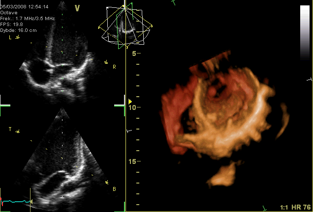
V Octave: This likely refers to the imaging modality or software used, indicating an advanced echocardiography system. “Octave” may signify its capability for detailed, high-resolution imaging.
Frek.: 1.7 MHZ/3.5 MHZ: This indicates the transducer frequency used for the ultrasound examination. Lower frequencies (e.g., 1.7 MHz) typically penetrate deeper, while higher frequencies (e.g., 3.5 MHz) offer better resolution for superficial structures.
FPS: 19.8: Stands for Frames Per Second, indicating the rate at which the ultrasound images are captured and displayed. A higher FPS allows for better visualization of rapid cardiac motion.
Dybde: 16.0 cm: This refers to the depth setting of the ultrasound scan, indicating that the machine is imaging structures up to 16.0 centimeters from the transducer. This parameter is adjusted based on the patient’s anatomy and the structures being examined.
HR 76: This is the heart rate, measured at 76 beats per minute. A normal heart rate is a key indicator of cardiac rhythm and overall cardiovascular health.
2D Echocardiogram Views (Upper and Middle Panels): These grayscale images represent standard two-dimensional ultrasound cross-sections of the heart. They allow for real-time visualization of cardiac chambers, valves, and wall motion, showing the heart’s pumping action.
3D Reconstruction (Right Panel and Lower Left Panel): This vividly colored image showcases a three-dimensional model of the heart, constructed from multiple 2D ultrasound slices. It provides a comprehensive spatial understanding of the heart’s external and internal structures, including the great vessels and chambers.
Echocardiography is a cornerstone of cardiovascular diagnostics, utilizing sound waves to create live images of the heart. This non-invasive technique allows clinicians to visualize the heart’s chambers, valves, great vessels, and pericardium, assessing their size, function, and blood flow patterns. The ability to acquire both 2D real-time images and construct detailed 3D models, as shown here, significantly enhances diagnostic accuracy and aids in the planning of medical and surgical interventions. This advanced imaging modality is particularly valuable for evaluating conditions affecting cardiac muscle, valve integrity, and the overall pumping efficiency of the heart.
The combination of 2D and 3D echocardiography provides a holistic view of cardiac health. The 2D views offer dynamic, cross-sectional insights into myocardial contractility, valve motion, and the presence of any abnormal fluid collections around the heart. The 3D reconstruction, on the other hand, allows for a comprehensive spatial understanding of complex cardiac anatomy, which is particularly useful for assessing congenital heart defects or guiding complex procedures. For example, assessing the heart rate (HR 76 bpm in this image) alongside visual data gives a complete picture of cardiac performance.
Advanced echocardiography plays a pivotal role in diagnosing a spectrum of cardiovascular diseases, from valvular heart disease and cardiomyopathy to congenital heart conditions and pericardial effusions. For instance, valvular heart disease, where heart valves are narrowed (stenosis) or leaky (regurgitation), can be precisely assessed using these techniques. Echocardiograms can visualize abnormal valve leaflet movement, measure blood flow velocities through valves using Doppler, and quantify the severity of regurgitation or stenosis, guiding treatment decisions.
- Echocardiography can detect abnormalities in heart muscle function, such as reduced ejection fraction, indicating heart failure.
- Doppler echocardiography measures blood flow velocity and direction, crucial for assessing valve function and shunt lesions.
- Transesophageal echocardiography (TEE) offers clearer images by placing the transducer closer to the heart.
- Stress echocardiography evaluates heart function during physical or pharmacological stress, detecting coronary artery disease.
This sophisticated imaging example highlights the indispensable role of modern echocardiography in contemporary cardiology. The ability to merge real-time functional assessment with detailed 3D anatomical visualization empowers cardiologists to make accurate diagnoses, monitor disease progression, and tailor treatment strategies effectively. This diagnostic power ultimately contributes to improved patient outcomes and a deeper understanding of the complexities of the human heart.

