This article explores the radiological appearance of deep vein thrombosis within the common iliac vein as seen on an abdominal computed tomography (CT) scan. We will examine the clinical significance of iliac vein thrombosis, the anatomy of the pelvic venous system, and the critical role of diagnostic imaging in preventing complications like pulmonary embolism.
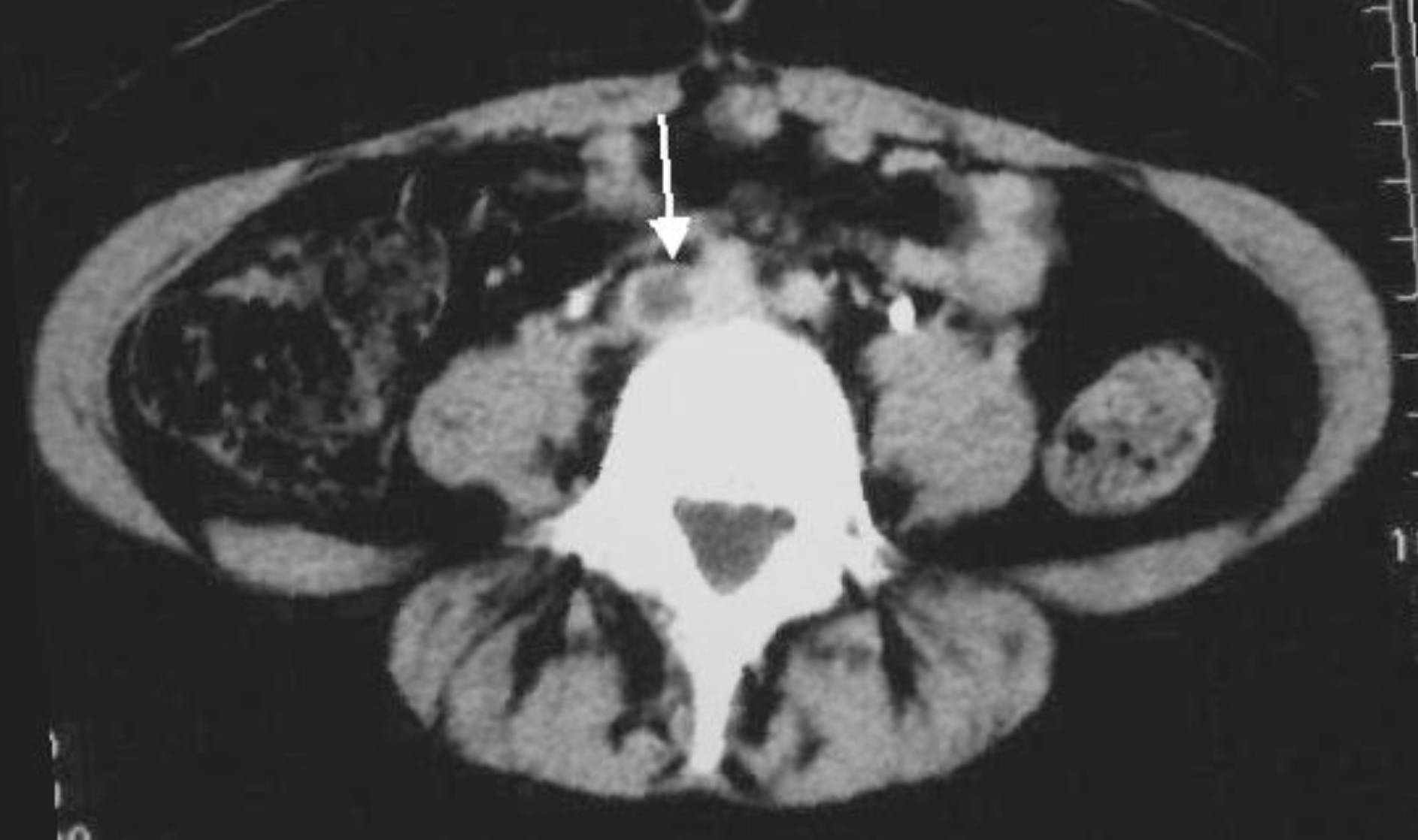
White arrow: The arrow points to a distinct filling defect located within the lumen of the right common iliac vein. Unlike the surrounding vessels which appear bright due to contrast enhancement, this area is dark and hypodense, indicating the presence of a solid thrombus (blood clot) obstructing normal blood flow.
Understanding Iliac Vein Thrombosis
Deep Vein Thrombosis (DVT) is a serious medical condition that typically occurs in the lower extremities, but it can extend proximally or originate in the large veins of the pelvis, known as the iliac veins. The image provided illustrates an axial CT slice of the abdomen, revealing a significant clot in the common iliac vein. Under normal circumstances, the iliac veins serve as the primary drainage pathways, carrying deoxygenated blood from the legs and pelvis into the inferior vena cava and back to the heart. When these vessels are obstructed, it creates a severe bottleneck in the venous system.
Diagnosing thrombosis in the pelvic veins presents a unique challenge compared to leg veins. While ultrasound is the gold standard for detecting clots in the thigh, sound waves often cannot penetrate deep enough to visualize the iliac veins due to interference from bowel gas and pelvic bone structure. Therefore, contrast-enhanced computed tomography (CT) venography is frequently utilized. As seen in the image, the CT scan allows for a clear cross-sectional view, differentiating between the patent (open) left iliac vein and the thrombosed (blocked) right iliac vein.
The clinical implications of an iliac vein DVT are generally more severe than those of distal clots. Because the iliac vein is a large-caliber vessel, the clot burden is substantial. This increases the risk of a massive pulmonary embolism if the clot dislodges. Furthermore, the complete occlusion of such a major vessel often leads to significant morbidity in the affected leg.
Common symptoms and risk factors associated with iliac vein thrombosis include:
- Severe, diffuse swelling of the entire leg and thigh.
- Pelvic pain or lower abdominal tenderness.
- Visible collateral veins on the lower abdomen or groin.
- Anatomical compression (such as May-Thurner syndrome).
- Prolonged immobilization or recent pelvic surgery.
Pathophysiology of Proximal DVT
Deep Vein Thrombosis involves the formation of a blood clot within the deep venous system. The formation of these clots is often described by Virchow’s Triad: stasis of blood flow, endothelial injury, and hypercoagulability. In the case of iliac vein thrombosis, anatomical factors often play a significant role. For example, May-Thurner syndrome is a condition where the left common iliac vein is compressed by the overlying right common iliac artery against the spine. This mechanical compression causes physical damage to the vein wall and slows blood flow, creating an ideal environment for a clot to form.
Once a thrombus forms in the iliac vein, it obstructs venous return. This leads to a rapid increase in venous pressure in the leg. Fluid leaks out of the capillaries and into the interstitial tissues, causing profound edema (swelling). In severe cases, the pressure can become so high that it compromises arterial blood flow into the leg, a condition known as phlegmasia cerulea dolens. This is a surgical emergency that can lead to gangrene and limb loss if the clot is not removed or dissolved immediately.
Diagnostic Imaging and Clinical Management
The abdominal CT scan is invaluable for assessment because it shows the “filling defect.” When contrast dye is injected into the bloodstream, it turns the liquid blood bright white on the scan. A clot, being a solid mass, does not absorb the dye and remains dark grey. In the provided image, the vertebral body (spine) is the bright white structure in the center, and the thrombosed vein is adjacent to it. Identifying this defect confirms the diagnosis and helps physicians measure the extent of the clot—specifically, whether it extends into the inferior vena cava.
Treatment for iliac vein thrombosis is aggressive. The primary goal is to prevent a pulmonary embolism, which occurs if the clot breaks loose and travels to the lungs. Standard therapy involves immediate anticoagulation with drugs like heparin, followed by oral anticoagulants. However, due to the size of iliac clots and the risk of post-thrombotic syndrome (chronic pain and swelling), interventional procedures are often considered. These may include catheter-directed thrombolysis, where “clot-busting” medication is delivered directly into the vein, or a mechanical thrombectomy to physically suck out the clot.
Conclusion
The detection of a common iliac vein thrombosis via CT imaging is a critical diagnostic finding that dictates immediate medical intervention. By visualizing the precise location and size of the occlusion, healthcare providers can prevent life-threatening complications and preserve the function of the lower limb. This image serves as a powerful reminder of the utility of advanced radiology in diagnosing complex vascular conditions that lie deep within the body, beyond the reach of a physical exam or standard ultrasound.

