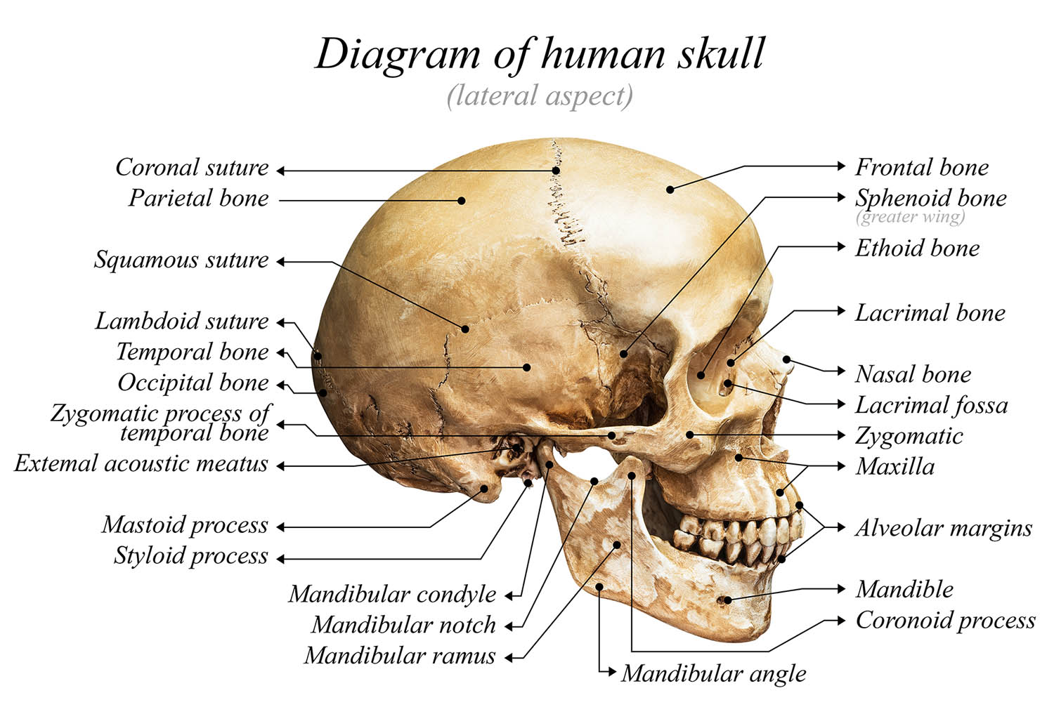
The cranial bones, forming the skull’s protective framework, are essential for safeguarding the brain and supporting facial structures, but their names and roles can challenge medical students. The mnemonic “PEST OF 6” simplifies memorization by organizing the six key cranial bones into a catchy, memorable phrase. This tool aids students in mastering skull anatomy for exams and clinical applications, such as interpreting head trauma imaging.
Mnemonic Statement
PEST OF 6
Mnemonic Breakdown
P
P stands for the parietal bones, paired structures forming the sides and roof of the cranium. These flat bones articulate at the sagittal suture, contributing to the skull’s strength and protecting the brain’s parietal lobes. Their broad surface is a common site for fractures in blunt trauma.
E
E represents the ethmoid, a single, delicate bone located at the skull’s midline, anterior to the sphenoid. It forms part of the nasal cavity and orbital walls, housing the cribriform plate critical for olfactory nerve transmission. Its fragility makes it susceptible to fractures in facial trauma.
S
S denotes the sphenoid, a complex, butterfly-shaped bone at the skull’s base, articulating with multiple cranial bones. It houses the pituitary gland in the sella turcica and forms part of the orbit, playing a key role in cranial stability. Sphenoid fractures can affect cranial nerves and require precise imaging.
T
T refers to the temporal bones, paired structures at the skull’s sides, housing the auditory and vestibular systems. They protect the middle and inner ear and anchor the mandible via the temporomandibular joint. Temporal bone fractures often result from lateral head impacts, risking hearing loss.
O
O stands for the occipital, a single bone forming the skull’s posterior base, cradling the cerebellum. It features the foramen magnum, allowing the spinal cord’s passage, and supports head movement via atlas articulation. Occipital fractures may disrupt critical neural pathways, necessitating urgent evaluation.
F
F represents the frontal, a single bone forming the forehead and anterior cranial vault, including the orbital roofs. It protects the frontal lobes and shapes facial appearance, with its sinuses aiding in voice resonance. Frontal bone fractures, common in high-impact injuries, often require surgical intervention.
Mnemonic Statement
PEST OF 6
Summary and Clinical Context
The “PEST OF 6” mnemonic streamlines the recall of the six cranial bones—parietal, ethmoid, sphenoid, temporal, occipital, and frontal—into a concise, vivid phrase that mirrors their anatomical significance. Its logical structure, emphasizing key bones in a memorable sequence, enables medical students to quickly retrieve this knowledge during high-pressure scenarios like exams or clinical rotations. By associating each letter with a bone’s unique role, the mnemonic fosters both rapid memorization and a deeper understanding of skull anatomy, essential for diagnosing conditions such as skull fractures or cranial nerve injuries.
In clinical practice, understanding cranial bone anatomy is critical for interpreting imaging studies, such as CT scans, used to assess head trauma. For instance, temporal bone fractures, accounting for roughly 20% of skull fractures, may disrupt auditory function, while ethmoid fractures can impair olfaction due to cribriform plate damage. The mnemonic’s simplicity aids students in pinpointing these bones’ locations and functions, enhancing diagnostic accuracy. Its utility extends to surgical planning, where precise knowledge of bones like the sphenoid, with its proximity to the pituitary, informs neurosurgical approaches.
Beyond academics, “PEST OF 6” supports interdisciplinary applications, benefiting radiologists, neurologists, and maxillofacial surgeons who rely on cranial bone familiarity for patient care. Students can amplify retention by pairing the mnemonic with visual aids, like 3D skull models, or integrating it with related mnemonics, such as those for facial bones, to build a robust anatomical framework. Its adaptability makes it valuable for beginners and advanced learners preparing for specialties like neurosurgery or otolaryngology, where cranial anatomy underpins clinical decision-making.
The mnemonic’s blend of memorability and practical relevance empowers students to excel academically and clinically. By embedding the cranial bones in an engaging phrase, it transforms a complex topic into an accessible, confidence-building tool. Whether studying for board exams or evaluating a patient with a suspected frontal bone fracture, “PEST OF 6” equips learners with the knowledge to navigate skull anatomy effectively.
Share Your Insights
How has “PEST OF 6” helped you master the cranial bones? Share your study tips or feedback below and explore our Carpal Bones Mnemonic for more anatomy resources!

