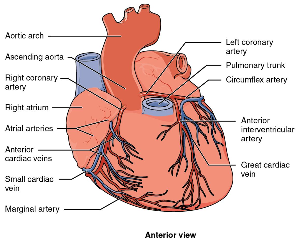
The coronary circulation is a vital network that delivers oxygen and nutrients to the heart muscle, and this image provides a detailed anterior view of its prominent surface vessels. By showcasing the arteries that sustain cardiac function, the diagram offers a clear perspective on their anatomical layout and importance to heart health. Exploring this illustration enhances understanding of the heart’s blood supply and its critical role in maintaining circulation.
Left coronary artery: The left coronary artery originates from the aorta and branches into the left anterior descending and circumflex arteries, supplying oxygenated blood to the left side of the heart. It plays a key role in nourishing the left ventricle and parts of the interventricular septum, supporting robust cardiac contraction.
Right coronary artery: The right coronary artery arises from the aorta and provides blood to the right atrium, right ventricle, and parts of the left ventricle via its branches. It is essential for maintaining the heart’s right-sided function and supporting electrical conduction through the atrioventricular node.
Left anterior descending artery: The left anterior descending artery, a major branch of the left coronary artery, runs down the anterior interventricular groove, delivering blood to the anterior wall of the left ventricle and the anterior septum. It is critical for the heart’s pumping efficiency and is a common site for coronary artery blockages.
Anatomical Structure of Coronary Circulation
The heart’s vascular network is intricately designed to support its continuous operation, and this anterior view highlights its key arteries. The diagram provides a focused look at the vessels that ensure myocardial health.
- The left coronary artery emerges near the aortic root, branching to cover the left heart.
- The right coronary artery supplies the right side, complementing the left coronary system.
- The left anterior descending artery targets the front of the left ventricle, a high-demand area.
- This layout reflects the heart’s need for a dual blood supply to maintain function.
The anterior view emphasizes the accessibility of these vessels for diagnostic imaging.
Functional Roles in Cardiac Perfusion
Coronary arteries deliver oxygen-rich blood during diastole, and this image illustrates their critical distribution. Their paths support the heart’s relentless workload across the anterior surface.
- The left coronary artery feeds the left ventricle, essential for systemic circulation.
- The right coronary artery sustains the right ventricle, aiding pulmonary circulation.
- The left anterior descending artery ensures the anterior wall’s perfusion, preventing ischemia.
- This coordinated supply maintains cardiac muscle vitality during each heartbeat.
Blockages can lead to severe outcomes like myocardial infarction.
Physical Characteristics and Clinical Relevance
The physical traits of these coronary arteries reflect their adaptation to the heart’s demands. Their structure influences their vulnerability to disease and treatment options.
- The left coronary artery’s larger size supports the left ventricle’s greater oxygen needs.
- The right coronary artery’s path varies, often determining coronary dominance.
- The left anterior descending artery’s straight course makes it prone to atherosclerotic plaques.
- Coronary angiography evaluates these vessels for stenosis, guiding interventions like stenting.
Their visibility on the anterior surface aids in surgical planning.
Importance in Cardiac Health
Preserving the patency of coronary circulation is crucial for long-term heart function. The health of these arteries directly impacts cardiac endurance and recovery.
- A healthy left coronary artery prevents left-sided heart failure risks.
- The right coronary artery’s integrity supports stable heart rhythms.
- The left anterior descending artery’s flow guards against anterior wall damage.
- Lifestyle changes, such as a low-cholesterol diet, protect these vessels from disease.
Regular stress tests monitor their condition effectively.
Conclusion
This diagram of coronary circulation from the anterior view provides a detailed illustration of the left coronary artery, right coronary artery, and left anterior descending artery, showcasing their roles in heart perfusion. By highlighting the vascular network that sustains the heart’s anterior surface, it underscores the importance of maintaining these arteries for optimal cardiac function. This understanding fosters greater awareness of coronary anatomy and the need to safeguard heart health through proactive care.

