The coronary circulation is a vital network of blood vessels that supplies oxygen and nutrients to the heart muscle, and this image showcases its prominent surface vessels from both anterior and posterior perspectives. With detailed views of the heart’s vascular anatomy, the diagram highlights the arteries responsible for sustaining cardiac function, offering a comprehensive look at their distribution. Exploring these illustrations provides valuable insights into the heart’s blood supply and its critical role in maintaining cardiovascular health.
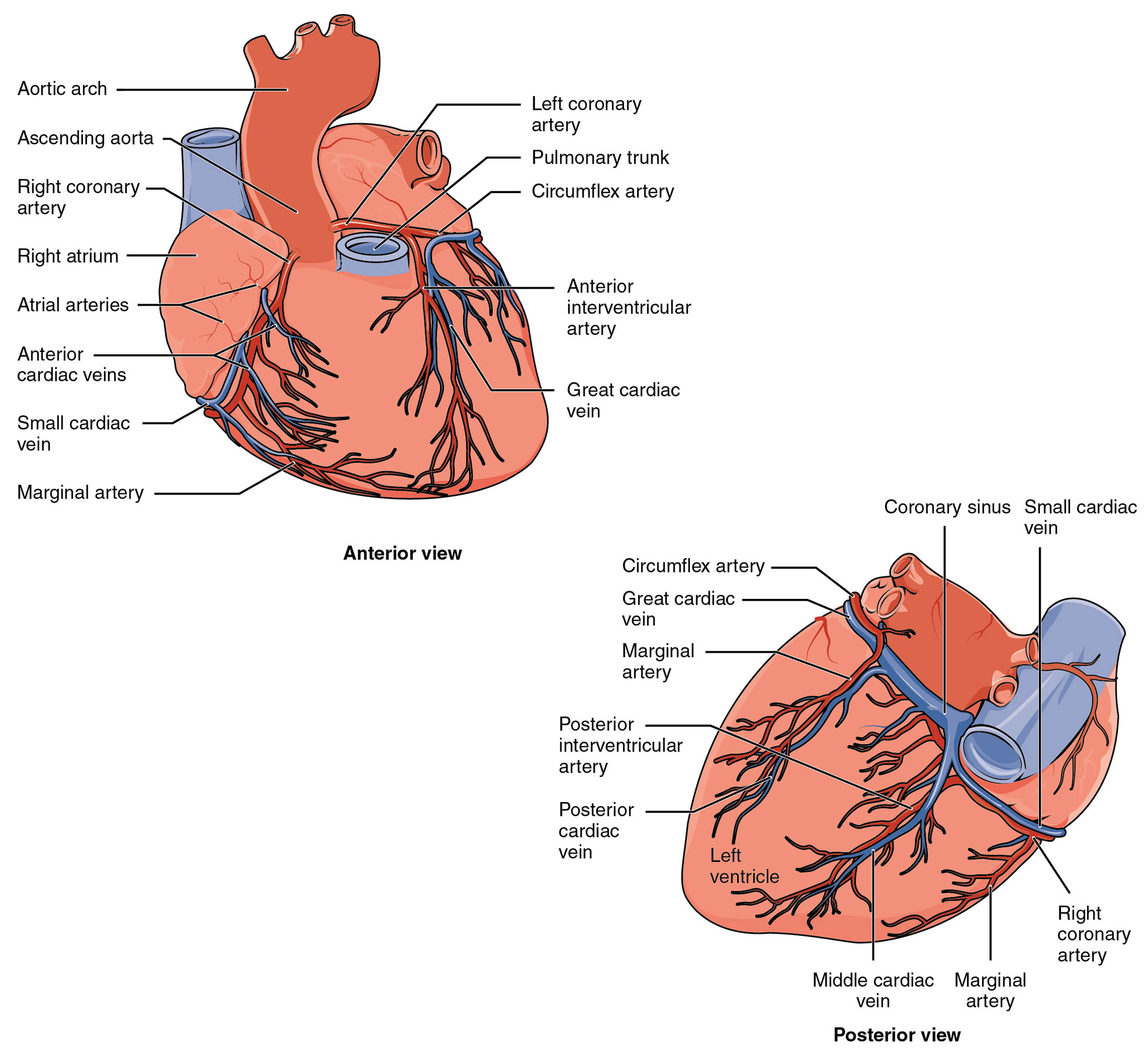
Left coronary artery: The left coronary artery originates from the aorta and branches into the left anterior descending and circumflex arteries, delivering oxygenated blood to the left side of the heart. It plays a crucial role in supplying the left ventricle and parts of the interventricular septum, ensuring robust cardiac contraction.
Right coronary artery: The right coronary artery arises from the aorta and supplies blood to the right atrium, right ventricle, and parts of the left ventricle via the posterior descending artery. It is essential for supporting the heart’s right side and maintaining electrical conduction through the atrioventricular node.
Circumflex artery: The circumflex artery, a branch of the left coronary artery, runs along the left atrioventricular groove, providing blood to the lateral wall of the left ventricle and the posterior surface. Its extensive reach ensures adequate perfusion to areas critical for systolic function.
Left anterior descending artery: The left anterior descending artery, a major branch of the left coronary artery, travels down the anterior interventricular groove, supplying blood to the anterior wall of the left ventricle and the anterior septum. It is vital for the heart’s pumping efficiency and is a common site for coronary artery disease.
Posterior descending artery: The posterior descending artery, often a branch of the right coronary artery, runs along the posterior interventricular groove, delivering blood to the posterior wall of both ventricles and the inferior septum. It supports the heart’s posterior region, contributing to overall cardiac stability.
Anatomical Structure of Coronary Circulation
The heart’s vascular network is intricately designed to nourish its muscular walls, and this diagram illustrates its layout from both views. These arteries form a critical lifeline for continuous cardiac operation.
- The left coronary artery emerges near the aortic root, branching to cover the left heart.
- The right coronary artery provides a parallel supply, nourishing the right heart and conduction system.
- The circumflex artery extends laterally, ensuring comprehensive left ventricular coverage.
- The left anterior descending artery and posterior descending artery target specific interventricular regions.
This dual-supply system enhances the heart’s resilience to localized blockages.
Functional Roles in Cardiac Perfusion
Coronary arteries deliver oxygen-rich blood during diastole, and this image highlights their functional distribution. Their precise paths support the heart’s relentless workload.
- The left coronary artery feeds the high-demand left ventricle, critical for systemic output.
- The right coronary artery sustains the right ventricle and supports rhythm regulation.
- The circumflex artery ensures lateral wall perfusion, preventing ischemic damage.
- The left anterior descending artery and posterior descending artery supply key septal and posterior areas.
Blockages in these arteries can lead to myocardial infarction if untreated.
Physical Characteristics and Clinical Relevance
The physical traits of these coronary arteries reflect their adaptation to the heart’s needs. Their structure influences susceptibility to disease and diagnostic approaches.
- The left coronary artery’s larger caliber supports the left ventricle’s greater workload.
- The right coronary artery’s path varies, often dominating in right-dominant hearts.
- The circumflex artery’s course makes it prone to occlusion, affecting lateral function.
- The left anterior descending artery and posterior descending artery are critical targets for angioplasty.
Coronary angiography assesses these vessels for stenosis or blockages.
Importance in Cardiac Health
Maintaining the patency of coronary circulation is essential for long-term heart function. These arteries’ health directly impacts cardiac endurance and recovery.
- A healthy left coronary artery prevents left-sided heart failure.
- The right coronary artery’s integrity supports stable heart rhythms.
- The circumflex artery’s perfusion reduces risk of lateral wall infarction.
- The left anterior descending artery and posterior descending artery’s flow guards against widespread damage.
Lifestyle changes and medical interventions like stents preserve their function.
Conclusion
This diagram of coronary circulation provides a detailed view of the left coronary artery, right coronary artery, circumflex artery, left anterior descending artery, and posterior descending artery, illustrating their roles in heart perfusion. By showcasing the anterior and posterior vascular networks, it emphasizes the heart’s reliance on these vessels for oxygen and nutrient delivery. This understanding fosters greater awareness of coronary anatomy and the importance of maintaining their health to support robust cardiac performance.

