Congenital heart defects are structural abnormalities present at birth that affect the heart’s functionality, often impacting blood flow and oxygenation. This diagram illustrates four common types—patent foramen ovale, coarctation of the aorta, patent ductus arteriosus, and tetralogy of Fallot—providing a visual guide to their anatomical differences. Understanding these defects through this image offers valuable insights into their diagnosis and management.
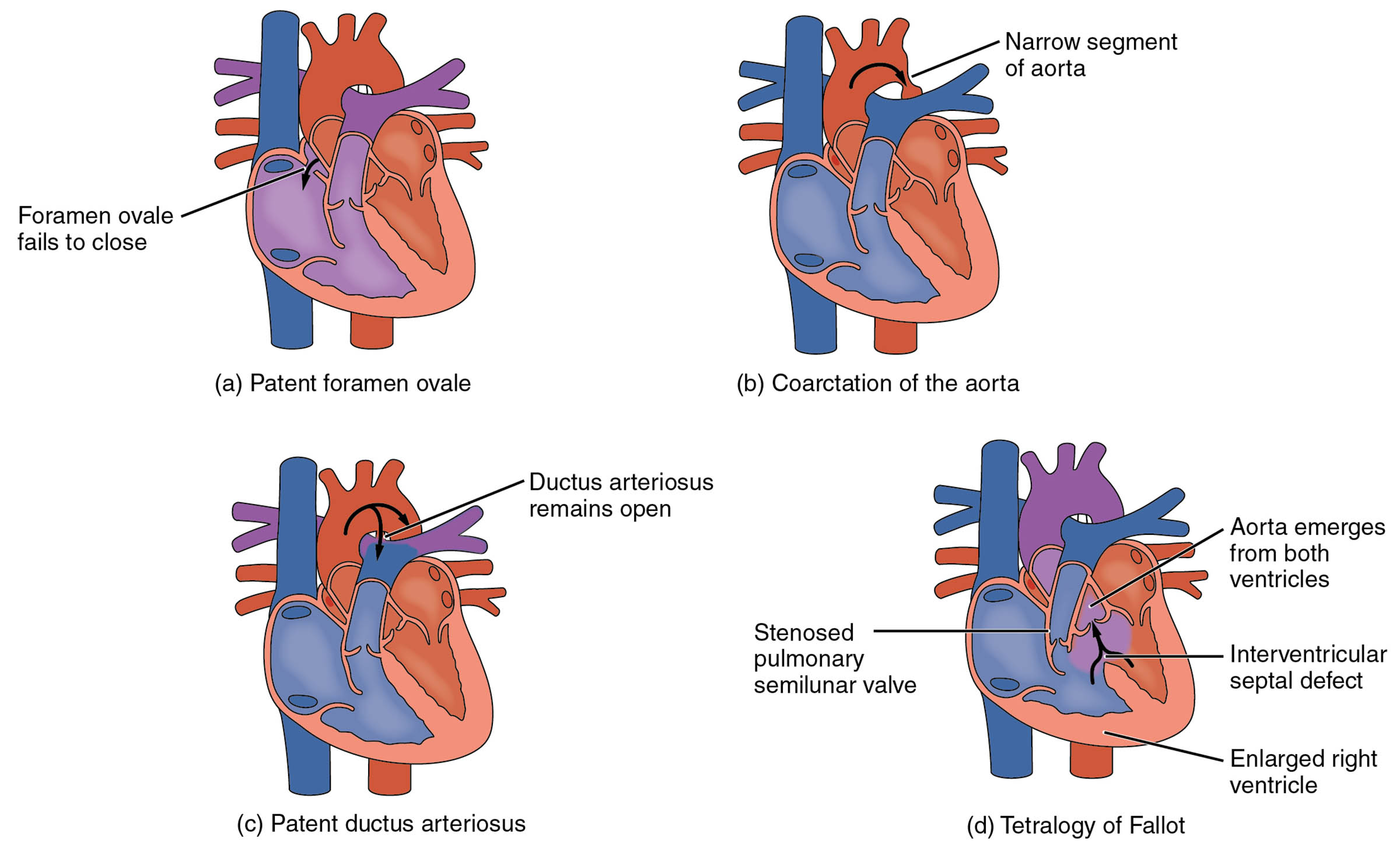
Foramen ovale fails to close: The foramen ovale fails to close when the flap between the atria does not seal after birth, allowing blood to flow between the right and left atria. This condition, known as patent foramen ovale, can lead to mixing of oxygenated and deoxygenated blood, potentially causing complications later in life.
Narrow segment of aorta: The narrow segment of aorta indicates a constriction in the aortic arch, characteristic of coarctation of the aorta, which restricts blood flow to the lower body. This narrowing increases pressure in the upper body and can lead to hypertension or heart failure if untreated.
Ductus arteriosus remains open: The ductus arteriosus remains open when the blood vessel connecting the pulmonary artery to the aorta fails to close after birth, a condition called patent ductus arteriosus. This persistent opening allows blood to bypass the lungs, potentially leading to increased workload on the heart.
Stenosed pulmonary valve semilunar valve: The stenosed pulmonary valve semilunar valve is a narrowed valve that obstructs blood flow from the right ventricle to the pulmonary artery, often seen in tetralogy of Fallot. This restriction reduces oxygen supply to the blood, contributing to cyanosis in affected individuals.
Aorta emerges from both ventricles: The aorta emerges from both ventricles due to an abnormal alignment in tetralogy of Fallot, where the aorta is positioned over the ventricular septal defect. This misplacement allows deoxygenated blood to enter the aorta, further complicating oxygenation.
Interventricular septal defect: The interventricular septal defect is a hole in the septum separating the ventricles, a key feature of tetralogy of Fallot, allowing mixing of oxygenated and deoxygenated blood. This defect increases the risk of heart strain and requires surgical correction.
Enlarged right ventricle: The enlarged right ventricle results from increased pressure and volume due to the defects in tetralogy of Fallot, such as the septal defect and pulmonary stenosis. This hypertrophy can lead to heart failure if not addressed through medical intervention.
Overview of Congenital Heart Defects
Congenital heart defects arise during fetal development, affecting the heart’s chambers, valves, or blood vessels. These illustrations provide a clear comparison of normal and abnormal heart structures.
- The foramen ovale fails to close, creating a persistent opening that can go unnoticed until adulthood.
- Narrow segment of aorta disrupts systemic circulation, often detectable through a weak pulse in the lower extremities.
- Ductus arteriosus remains open leads to a characteristic machinery-like heart murmur.
- Stenosed pulmonary valve semilunar valve and interventricular septal defect in tetralogy of Fallot contribute to reduced oxygen levels.
Early detection through echocardiography is critical for effective management.
Anatomical and Physiological Impact
These defects alter the heart’s normal anatomy, impacting blood flow and oxygenation. The diagrams highlight the specific changes associated with each condition.
- Foramen ovale fails to close allows right-to-left shunting, which may increase stroke risk in some cases.
- Narrow segment of aorta elevates blood pressure above the constriction, straining the heart.
- Ductus arteriosus remains open causes a left-to-right shunt, overloading the pulmonary circulation.
- Aorta emerges from both ventricles and enlarged right ventricle reflect the complex interplay of defects in tetralogy of Fallot.
These alterations can lead to symptoms like cyanosis, fatigue, or poor growth in infants.
Causes and Risk Factors
The development of congenital heart defects involves genetic and environmental influences. Identifying these factors aids in prevention and counseling.
- Genetic mutations, such as those in the 22q11.2 deletion syndrome, increase the likelihood of defects like tetralogy of Fallot.
- Maternal factors, including diabetes or rubella infection during pregnancy, contribute to fetal heart abnormalities.
- Folic acid deficiency has been linked to neural tube defects but may also play a role in heart development.
- Prenatal screening, including fetal ultrasound, helps identify defects early for planned interventions.
Awareness of these risks supports better prenatal care.
Treatment and Management
Managing congenital heart defects requires tailored approaches based on the specific defect. Treatment options range from monitoring to surgical correction.
- Patent foramen ovale may close spontaneously or require catheter-based closure if symptomatic.
- Coarctation of the aorta often necessitates surgical repair or balloon angioplasty to widen the artery.
- Patent ductus arteriosus can be treated with medication like indomethacin or surgical ligation.
- Tetralogy of Fallot requires open-heart surgery to correct the interventricular septal defect and relieve pulmonary stenosis.
Long-term follow-up ensures optimal heart function and quality of life.
Conclusion
This diagram of congenital heart defects—including patent foramen ovale, coarctation of the aorta, patent ductus arteriosus, and tetralogy of Fallot—offers a detailed visual representation of these conditions. By illustrating the foramen ovale fails to close, narrow segment of aorta, ductus arteriosus remains open, stenosed pulmonary valve semilunar valve, aorta emerges from both ventricles, interventricular septal defect, and enlarged right ventricle, it provides a foundation for understanding their impact. This knowledge empowers individuals to recognize symptoms and seek timely medical care for effective management.

