Tetralogy of Fallot is a complex congenital heart defect characterized by an abnormal opening in the interventricular septum, leading to significant circulatory challenges. This diagram illustrates the key anatomical features of this condition, including the ventricular septal defect and associated abnormalities, providing a visual aid to understand its impact on heart function. Exploring this image offers critical insights into the condition’s structure and the importance of timely medical intervention.
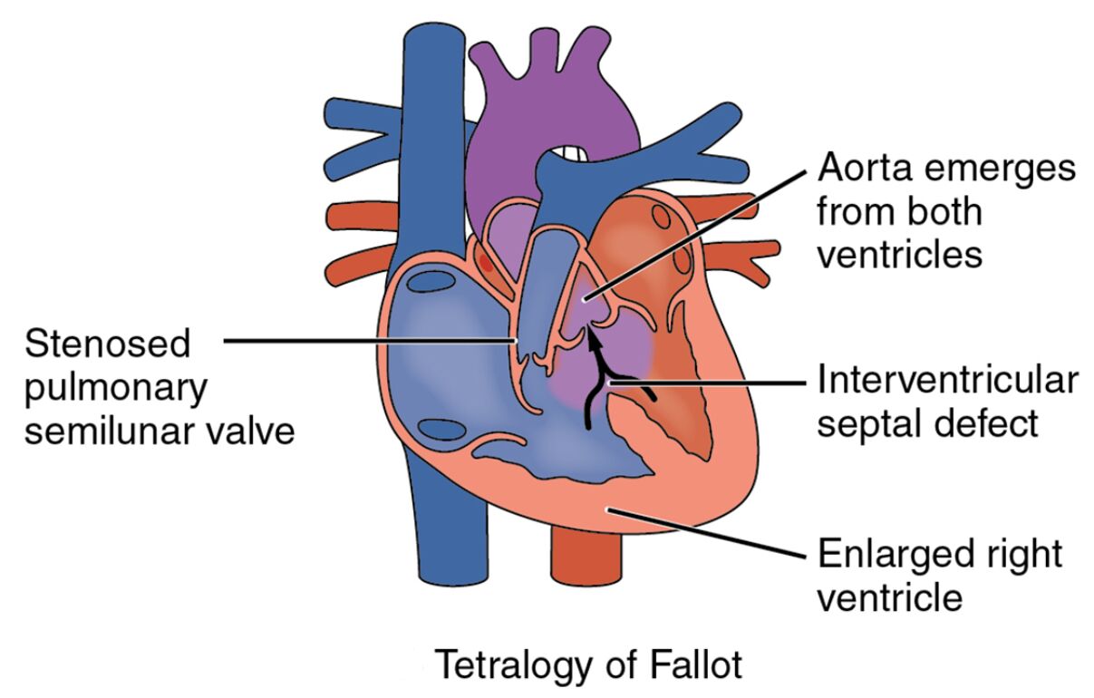
Right ventricle: The right ventricle pumps deoxygenated blood into the pulmonary artery, but in tetralogy of Fallot, it may become hypertrophied due to increased pressure from pulmonary stenosis. This chamber’s altered function contributes to reduced blood flow to the lungs, leading to cyanosis.
Left ventricle: The left ventricle pumps oxygenated blood into the aorta for systemic circulation, maintaining normal pressure and output. In tetralogy of Fallot, its function may be indirectly affected by the right ventricle’s overload and the mixing of blood.
Interventricular septal defect: The interventricular septal defect is an abnormal hole in the wall separating the ventricles, allowing oxygenated and deoxygenated blood to mix. This defect increases the workload on the heart and reduces oxygen saturation in the systemic circulation.
Overriding aorta: The overriding aorta is positioned abnormally over the ventricular septal defect, receiving blood from both ventricles instead of just the left. This misalignment contributes to the delivery of deoxygenated blood to the body, exacerbating cyanosis.
Pulmonary stenosis: Pulmonary stenosis is a narrowing of the pulmonary valve or artery, obstructing blood flow from the right ventricle to the lungs. This restriction forces more blood through the septal defect and overriding aorta, worsening oxygen deficiency.
Anatomical Overview of Tetralogy of Fallot
Tetralogy of Fallot involves multiple structural abnormalities within the heart, and this diagram provides a detailed view of its components. Recognizing these features is essential for understanding the condition’s complexity.
- The right ventricle adapts to the obstruction with thickened walls, reflecting its increased effort.
- The left ventricle remains relatively unaffected structurally but supports the altered circulation.
- The interventricular septal defect serves as a pathway for blood mixing, a hallmark of this defect.
- The overriding aorta and pulmonary stenosis together create a unique circulatory pattern.
This combination of defects is one of the most common causes of blue baby syndrome.
Physiological Impact and Symptoms
The abnormalities in tetralogy of Fallot significantly alter blood flow and oxygenation, leading to recognizable symptoms. The diagram highlights the functional consequences of these changes.
- The right ventricle’s hypertrophy increases the risk of heart failure due to sustained pressure.
- The interventricular septal defect allows deoxygenated blood to enter the overriding aorta, causing cyanosis.
- Pulmonary stenosis limits lung blood flow, forcing the heart to compensate with shunting.
- Symptoms include difficulty feeding, clubbing of fingers, and episodes of severe cyanosis known as “tet spells.”
Echocardiography is critical for confirming the diagnosis and assessing severity.
Causes and Risk Factors
The development of tetralogy of Fallot involves genetic and environmental influences during fetal development. Understanding these factors aids in prevention and early detection.
- Genetic syndromes like DiGeorge syndrome are linked to a higher incidence of this defect.
- Maternal factors, such as phenylketonuria or viral infections, can contribute to congenital heart issues.
- Chromosomal abnormalities, including trisomy 21, increase the likelihood of heart defects.
- Prenatal ultrasound can identify structural anomalies, enabling early planning for care.
Folic acid supplementation during pregnancy may help reduce related risks.
Diagnosis and Treatment Options
Diagnosing and managing tetralogy of Fallot requires a multidisciplinary approach based on the defect’s impact. Imaging and clinical evaluation guide treatment decisions.
- Echocardiography visualizes the interventricular septal defect and pulmonary stenosis for accurate diagnosis.
- Initial management may include prostaglandin E1 to maintain ductus arteriosus patency in neonates.
- Surgical repair, often involving patch closure of the interventricular septal defect, is the definitive treatment.
- Follow-up care monitors for residual issues like pulmonary regurgitation or right ventricle strain.
Timely intervention significantly improves long-term outcomes.
Clinical Relevance and Long-Term Outlook
Understanding the implications of tetralogy of Fallot is vital for long-term health management. The condition’s effects depend on the severity of the defects and surgical success.
- The right ventricle’s health post-surgery determines exercise tolerance and quality of life.
- The overriding aorta’s alignment may require ongoing assessment to prevent aortic root dilation.
- Correcting the pulmonary stenosis and interventricular septal defect reduces cyanosis risk.
- Adults with repaired tetralogy need regular cardiology follow-ups to monitor heart function.
Lifestyle adjustments, like avoiding strenuous activity initially, support recovery.
Conclusion
This diagram of tetralogy of Fallot provides a detailed illustration of the right ventricle, left ventricle, interventricular septal defect, overriding aorta, and pulmonary stenosis, showcasing the impact of this congenital heart defect. By highlighting how these abnormalities affect blood oxygenation and circulation, it underscores the need for early diagnosis and surgical correction. This knowledge equips individuals with the tools to manage the condition effectively, enhancing overall health and well-being.

