Patent foramen ovale (PFO) is a congenital heart defect characterized by an abnormal opening in the interatrial septum, often due to the failure of the foramen ovale to close after birth. This diagram provides a clear visual representation of the heart’s anatomy, highlighting the location and impact of this defect on blood flow between the atria. Exploring this image offers valuable insights into the condition’s implications and its relevance to cardiovascular health.
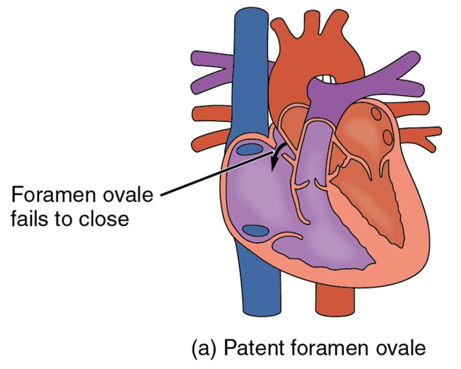
Right atrium: The right atrium receives deoxygenated blood from the body via the superior and inferior vena cava, preparing it for passage to the right ventricle. In a patent foramen ovale, this chamber may allow blood to shunt into the left atrium, altering normal circulation.
Left atrium: The left atrium collects oxygenated blood from the lungs through the pulmonary veins, directing it to the left ventricle for systemic circulation. A patent foramen ovale can cause deoxygenated blood to mix with this oxygenated blood, potentially affecting oxygen delivery.
Interatrial septum: The interatrial septum is the wall separating the right and left atria, normally closing the foramen ovale after birth to prevent blood mixing. In patent foramen ovale, this septum contains an incomplete closure, leading to an abnormal opening.
Foramen ovale: The foramen ovale is a fetal opening in the interatrial septum that allows blood to bypass the lungs during prenatal development, typically closing shortly after birth. When it fails to close, it results in a patent foramen ovale, permitting ongoing shunting of blood.
Anatomical Overview of Patent Foramen Ovale
The heart’s atrial septum plays a critical role in maintaining proper blood flow, and this diagram illustrates its normal and defective states. Understanding the anatomy helps in recognizing the impact of this congenital defect.
- The right atrium serves as the entry point for deoxygenated blood, which should flow only to the right ventricle.
- The left atrium is designed to handle oxygenated blood, ensuring efficient distribution to the body.
- The interatrial septum forms a barrier that, when intact, prevents mixing of blood between the atria.
- The foramen ovale’s persistence creates a pathway for blood to move abnormally, altering cardiac dynamics.
This defect is present in about 20-25% of the population, often remaining asymptomatic.
Physiological Impact and Symptoms
Patent foramen ovale affects blood flow patterns, potentially leading to noticeable symptoms in some cases. The diagram highlights how this defect influences cardiac function.
- Right atrium blood may shunt into the left atrium, reducing oxygen saturation in systemic circulation.
- This mixing can lead to paradoxical embolism, where clots travel to the brain, causing stroke.
- The interatrial septum’s incomplete closure may go unnoticed unless significant shunting occurs.
- Symptoms like migraines or fatigue may arise, though many individuals remain asymptomatic.
Echocardiography with bubble study is used to diagnose this condition accurately.
Causes and Risk Factors
The development of patent foramen ovale involves genetic and developmental factors during fetal growth. Identifying these aids in understanding its prevalence.
- Genetic predisposition can influence septal development, increasing the likelihood of the foramen ovale remaining open.
- Maternal factors, such as diabetes or obesity, may contribute to congenital heart defects.
- The condition is more common in individuals with a family history of cardiac anomalies.
- Prenatal ultrasound can detect septal issues, guiding postnatal care planning.
Folic acid supplementation during pregnancy may reduce related congenital risks.
Diagnosis and Treatment Options
Diagnosing and managing patent foramen ovale requires a tailored approach based on clinical presentation. Advanced imaging plays a key role in assessment.
- Transesophageal echocardiography visualizes the interatrial septum to confirm the defect’s presence.
- Asymptomatic cases may only need monitoring, avoiding invasive procedures.
- Catheter-based closure is an option for those with recurrent strokes linked to the foramen ovale.
- Medications like anticoagulants manage clot risk if closure isn’t pursued.
Regular follow-ups ensure the condition does not progress to more serious complications.
Clinical Relevance and Long-Term Outlook
Understanding the implications of patent foramen ovale is essential for long-term health management. The defect’s impact varies widely among individuals.
- The right atrium’s shunting can increase stroke risk, particularly during activities that raise right heart pressure.
- The left atrium’s exposure to deoxygenated blood may subtly affect exercise tolerance over time.
- The interatrial septum’s condition can be monitored with periodic imaging to assess changes.
- Most individuals with an isolated patent foramen ovale lead normal lives with proper care.
Lifestyle adjustments, like avoiding scuba diving, may be recommended for at-risk cases.
Conclusion
This diagram of patent foramen ovale provides a detailed view of the right atrium, left atrium, interatrial septum, and foramen ovale, illustrating the heart’s response to this congenital defect. By showing how the failure of the foramen ovale to close affects blood flow, it underscores the importance of early detection and management. This understanding equips individuals with the knowledge to address potential health challenges associated with this condition effectively.

