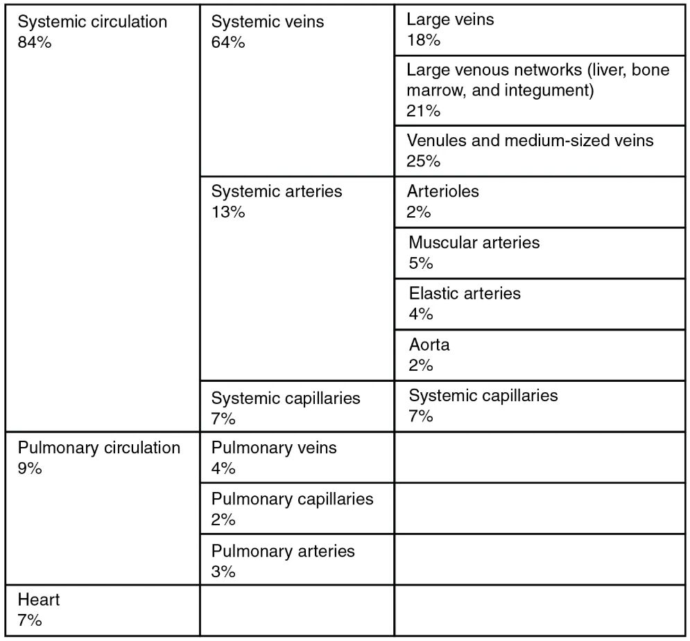
The human circulatory system plays a vital role in maintaining homeostasis by transporting oxygen, nutrients, hormones, and waste products throughout the body. This distribution of blood flow chart illustrates how blood volume is allocated across various components of the systemic and pulmonary circulations, as well as the heart, providing essential insights into cardiovascular physiology and its implications for health and medical practice.
Systemic circulation Systemic circulation refers to the pathway that carries oxygenated blood from the heart to the body’s tissues and returns deoxygenated blood back to the heart. It accounts for the majority of blood volume, ensuring that organs and tissues receive necessary nutrients while removing metabolic wastes.
84% This percentage represents the proportion of total blood volume dedicated to the systemic circulation, highlighting its dominance in the overall circulatory system. Such a high allocation underscores the extensive network required to support the body’s metabolic demands.
Systemic veins Systemic veins are the vessels that collect deoxygenated blood from the body’s tissues and transport it back to the heart. They form a crucial part of the return pathway in circulation, accommodating a significant portion of blood volume due to their capacitance function.
64% The 64% figure indicates the substantial blood volume held within systemic veins, reflecting their role as blood reservoirs. This high percentage allows for flexibility in blood distribution during varying physiological states, such as exercise or rest.
Large veins Large veins include major vessels like the superior and inferior vena cava, which drain blood from the upper and lower body, respectively. They are designed to handle high volumes of blood with minimal resistance, facilitating efficient return to the heart.
18% 18% of blood volume is contained in large veins, emphasizing their capacity to store blood temporarily. This allocation helps in maintaining venous return and stabilizing blood pressure.
Large venous networks (liver, bone marrow, and integument) Large venous networks in areas like the liver, bone marrow, and integument (skin) serve as specialized reservoirs that can release or store blood as needed. These networks support organ-specific functions, such as detoxification in the liver and hematopoiesis in the bone marrow.
21% The 21% allocation to large venous networks illustrates their importance in buffering blood volume changes. This percentage ensures these vital areas have adequate perfusion for their metabolic activities.
Venules and medium-sized veins Venules and medium-sized veins bridge the gap between capillaries and larger veins, collecting blood from microvascular beds. They play a key role in regulating blood flow through constriction and dilation, influenced by local metabolic needs.
25% 25% of blood is held in venules and medium-sized veins, allowing for fine-tuned control of venous return. This volume supports the transition from tissue-level exchange to central circulation.
Systemic arteries Systemic arteries carry oxygenated blood away from the heart to the tissues, branching into progressively smaller vessels. They maintain high pressure to propel blood throughout the body, with elastic properties to accommodate cardiac output.
13% The 13% blood volume in systemic arteries reflects the pressurized nature of arterial flow. This allocation is critical for delivering oxygen efficiently to distant organs.
Arterioles Arterioles are small arteries that regulate blood flow into capillary beds through smooth muscle contraction. They act as resistance vessels, controlling tissue perfusion and contributing to blood pressure regulation.
2% Only 2% of blood is in arterioles, but this small volume has a disproportionate impact on vascular resistance. Their ability to adjust diameter allows precise distribution based on tissue demand.
Muscular arteries Muscular arteries, also known as distributing arteries, have thick muscular walls to direct blood to specific regions. They distribute blood to organs and can vasoconstrict or vasodilate to meet varying needs.
5% 5% blood volume in muscular arteries supports their role in active distribution. This percentage enables rapid adjustments during physical activity or stress.
Elastic arteries Elastic arteries, such as the aorta and its major branches, contain elastic fibers to absorb systolic pressure and maintain continuous flow. They function as a Windkessel, smoothing out pulsatile blood flow from the heart.
4% The 4% allocation to elastic arteries highlights their buffering capacity. This volume helps in sustaining diastolic pressure for coronary and cerebral perfusion.
Aorta The aorta is the largest artery, originating from the left ventricle and branching to supply the entire body. It withstands the highest pressures and distributes blood to all systemic branches.
2% 2% of blood in the aorta underscores its central role in initial propulsion. This small but critical volume ensures immediate distribution post-ejection.
Systemic capillaries Systemic capillaries are the site of nutrient and gas exchange between blood and tissues. Their thin walls facilitate diffusion, making them essential for cellular metabolism.
7% 7% blood volume in systemic capillaries facilitates ongoing exchange processes. This percentage allows for sufficient contact time between blood and tissues.
Pulmonary circulation Pulmonary circulation transports deoxygenated blood from the heart to the lungs for oxygenation and returns oxygenated blood back. It operates at lower pressure compared to systemic circulation, focusing on gas exchange.
9% The 9% allocation to pulmonary circulation emphasizes its specialized function in respiration. This volume supports efficient oxygenation without overburdening the right heart.
Pulmonary veins Pulmonary veins carry oxygenated blood from the lungs to the left atrium. Unlike systemic veins, they transport oxygen-rich blood, completing the pulmonary loop.
4% 4% blood in pulmonary veins ensures swift return of oxygenated blood. This percentage maintains the flow for systemic distribution.
Pulmonary capillaries Pulmonary capillaries surround alveoli in the lungs, enabling gas exchange. They have a vast surface area for rapid diffusion of oxygen and carbon dioxide.
2% The modest 2% in pulmonary capillaries is optimized for quick transit. This allocation maximizes exposure to alveolar air.
Pulmonary arteries Pulmonary arteries deliver deoxygenated blood from the right ventricle to the lungs. They branch extensively to perfuse lung tissue uniformly.
3% 3% blood volume in pulmonary arteries supports low-resistance flow. This percentage aids in preventing right ventricular strain.
Heart The heart is the muscular pump driving circulation, with chambers that fill and eject blood rhythmically. It requires its own coronary supply to function continuously.
7% 7% of blood within the heart reflects its role as a dynamic reservoir. This volume is essential for cardiac output maintenance.
Overview of Blood Flow Distribution
The chart provides a snapshot of how blood is distributed at rest, offering a foundation for understanding cardiovascular dynamics. This knowledge is crucial for interpreting physiological responses and pathological conditions.
- Blood distribution is not static; it adjusts based on factors like activity level, hydration, and hormonal influences.
- The systemic component dominates because it services the entire body, while pulmonary focuses solely on lungs.
- Percentages add up to 100%, representing total blood volume, typically around 5 liters in adults.
- Variations can occur in conditions like shock or heart failure, altering these proportions.
Importance of Systemic Circulation
Systemic circulation ensures that every cell receives vital substances for survival. It integrates with other systems, such as endocrine and immune, to maintain overall health.
- Veins hold the largest share due to their compliance, acting as a capacitance system to buffer volume changes.
- Arteries, though smaller in volume, generate the pressure gradient needed for flow.
- Capillaries, with their 7% allocation, are where microcirculation occurs, involving processes like filtration and reabsorption per Starling’s forces.
- Hormones like vasopressin and aldosterone influence venous tone, affecting distribution.
Breakdown of Systemic Veins
Veins in the systemic circuit are categorized by size and function, each contributing uniquely to blood return. Their walls are thinner than arteries, relying on valves and muscle pumps for flow.
- Large veins, at 18%, include the venae cavae, providing low-resistance pathways.
- Large venous networks, comprising 21%, in organs like the liver (portal system) store blood and support detoxification.
- Venules (part of the 25% with medium-sized veins) drain capillaries, while medium-sized veins converge flows from regions like limbs.
- These vessels can dilate to hold more blood during rest or constrict during exercise to enhance return.
Breakdown of Systemic Arteries
Arteries progressively decrease in size from the aorta outward, each type adapted for specific roles. Their elastic and muscular components allow for pulse dampening and flow regulation.
- The aorta, at 2%, is the root of the arterial tree, with thick walls to handle ejection forces.
- Elastic arteries (4%) recoil to propel blood forward during diastole.
- Muscular arteries (5%) distribute to organs, controlled by sympathetic innervation.
- Arterioles (2%) are key in autoregulation, responding to local metabolites like adenosine.
Role of Pulmonary Circulation
Pulmonary circulation operates in parallel with systemic, optimizing gas exchange. It receives the entire cardiac output from the right ventricle, ensuring blood is refreshed continuously.
- Pulmonary arteries (3%) carry deoxygenated blood, branching into lobar and segmental vessels.
- Capillaries (2%) form a dense network around alveoli, where hematocrit may increase due to plasma filtration.
- Veins (4%) converge to four main trunks entering the left atrium.
- This circuit’s low pressure (about 15 mmHg mean) prevents fluid leakage into lung tissue.
The Heart’s Contribution
The heart, while pumping, holds 7% of blood volume in its chambers. This intracardiac blood is crucial for preload and stroke volume per Frank-Starling mechanism.
- Atria act as primers, ventricles as powerhouses.
- Coronary circulation, though not detailed here, perfuses the myocardium.
- Electrocardiographic synchronization ensures efficient filling and emptying.
- Pathologies like valvular disease can disrupt this balance.
In summary, this blood flow distribution chart reveals the intricate balance within the circulatory system, where each component’s allocation supports life-sustaining functions. Appreciating these proportions enhances comprehension of how the body adapts to demands, from daily activities to medical interventions, fostering better approaches to cardiovascular care.

