The anatomy of the human neck is a complex network of muscles, glands, and vital neurovascular bundles that facilitate essential physiological functions. This detailed cadaveric dissection highlights the common carotid artery and its relationship to the scalene muscles, brachial plexus, and various strap muscles, providing a foundational understanding for clinical practice and surgical interventions. Mastery of these landmarks is crucial for professionals performing procedures such as carotid endarterectomy, nerve blocks, or central venous access.
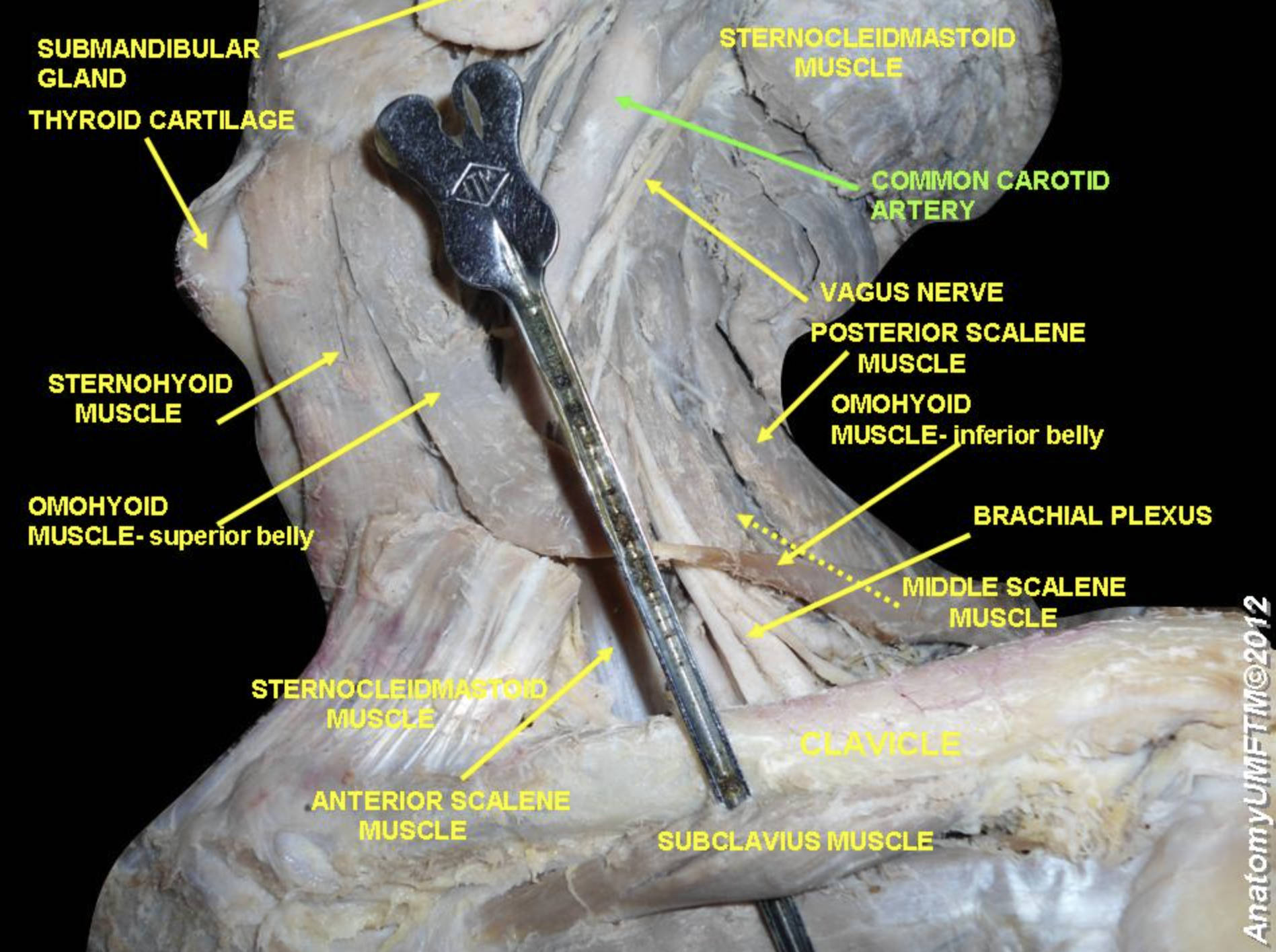
SUBMANDIBULAR GLAND: This is a major salivary gland located in the submandibular triangle, just beneath the body of the mandible. It plays a crucial role in oral health by secreting saliva through Wharton’s duct to aid in digestion and mucosal protection.
THYROID CARTILAGE: Often referred to as the “Adam’s apple,” this is the largest of the nine cartilages that compose the laryngeal skeleton. It serves as a protective shield for the vocal cords and provides an attachment point for various laryngeal and pharyngeal muscles.
STERNOHYOID MUSCLE: A thin, narrow muscle belonging to the infrahyoid group that connects the sternum and the clavicle to the hyoid bone. Its primary action is to depress the hyoid bone during the final stages of swallowing and speech.
OMOHYOID MUSCLE- superior belly: This portion of the omohyoid muscle originates from an intermediate tendon and attaches to the lower border of the hyoid bone. Working with the inferior belly, it acts to depress the hyoid bone and tense the deep cervical fascia to prevent venous collapse.
STERNOCLEIDOMASTOID MUSCLE: This robust, superficial muscle is a primary landmark of the neck, running from the sternum and clavicle to the mastoid process. It is responsible for rotating the head to the opposite side and flexing the neck when acting bilaterally.
ANTERIOR SCALENE MUSCLE: This muscle arises from the transverse processes of the C3-C6 vertebrae and inserts onto the scalene tubercle of the first rib. It is an essential anatomical landmark as it separates the subclavian vein from the subclavian artery and the brachial plexus.
SUBCLAVIUS MUSCLE: A small, triangular muscle situated in the space between the first rib and the clavicle. It functions to stabilize the clavicle during movements of the shoulder girdle and offers a layer of protection to the underlying neurovascular structures.
CLAVICLE: Commonly known as the collarbone, this long bone serves as a strut between the scapula and the sternum to support the upper limb. It is the only long bone in the human body that lies horizontally and is frequently used as a landmark for regional anesthesia.
MIDDLE SCALENE MUSCLE: This is the largest and longest of the three scalene muscles, originating from the C2-C7 vertebrae and inserting onto the first rib. It primarily functions to elevate the first rib during forced inspiration and aids in lateral neck flexion.
BRACHIAL PLEXUS: A complex network of nerves formed by the anterior rami of the C5-T1 spinal nerves that provides motor and sensory innervation to the upper limb. It emerges through the interscalene space, located between the anterior and middle scalene muscles.
OMOHYOID MUSCLE- inferior belly: This segment arises from the superior border of the scapula and travels obliquely across the lower neck before joining the superior belly via an intermediate tendon. It serves to divide the posterior triangle of the neck into the occipital and the supraclavicular triangle.
POSTERIOR SCALENE MUSCLE: The smallest and deepest of the scalene muscles, it originates from the lower cervical vertebrae and inserts onto the second rib. Its contraction assists in elevating the second rib during breathing and tilting the neck laterally.
VAGUS NERVE: Known as the tenth cranial nerve (CN X), it is the longest cranial nerve and carries extensive parasympathetic fibers to the thorax and abdomen. In the neck, it descends within the carotid sheath, positioned between the internal jugular vein and the common carotid artery.
COMMON CAROTID ARTERY: This major vessel provides the primary oxygenated blood supply to the head and neck. It ascends through the neck and typically bifurcates into the internal and external carotid arteries at the level of the upper border of the thyroid cartilage, a point known as the carotid bifurcation.
Structural Organization of the Neck and Carotid System
The neck functions as a vital transition zone between the head, the thorax, and the upper limbs. Within this relatively small space, critical life-sustaining structures are tightly packed and organized into compartments by layers of deep cervical fascia. The most prominent of these structures is the carotid system, which is housed within a tubular investment of fascia. This organization protects the high-pressure arterial system while allowing for the necessary flexibility and movement of the cervical spine.
Understanding the spatial relationships of the neck is fundamental for identifying clinical pathology. For example, the interscalene space is a primary site for administering nerve blocks for shoulder surgery, but its proximity to the dome of the pleura and the phrenic nerve requires precise anatomical knowledge. Furthermore, the strap muscles, such as the sternohyoid and omohyoid, are often retracted or divided during thyroid surgery to gain access to the underlying gland.
Key anatomical groupings highlighted in this view include:
- The scalene muscles (anterior, middle, and posterior), which act as accessory muscles of respiration.
- The infrahyoid “strap” muscles, which manage the positioning of the hyoid bone and larynx.
- The major neurovascular bundle, consisting of the common carotid artery, vagus nerve, and internal jugular vein.
- The brachial plexus, which serves as the nervous highway to the arm and hand.
Physiological Roles and Clinical Significance
The physiological importance of the common carotid artery cannot be overstated, as it is the conduit for blood flow to the brain. The carotid sinus, located at the base of the internal carotid artery near the bifurcation, contains baroreceptors that monitor systemic blood pressure. These receptors send signals via the glossopharyngeal nerve to the brainstem to regulate heart rate and vascular resistance, maintaining homeostatic balance. Any disruption in this area, such as the buildup of atherosclerotic plaque, can lead to carotid artery stenosis, significantly increasing the risk of ischemic stroke.
The vagus nerve provides another layer of physiological regulation, carrying sensory information from the aortic arch baroreceptors and chemoreceptors. It also provides the parasympathetic “rest and digest” signals that slow the heart rate and stimulate digestive processes. Because the vagus nerve lies in such close proximity to the carotid artery, surgeons must exercise extreme caution during neck dissections to avoid accidental injury, which could result in vocal cord paralysis or autonomic dysfunction.
Furthermore, the muscles of the neck, particularly the sternocleidomastoid and the scalenes, are not only involved in movement but also serve as indicators of respiratory distress. When a patient is struggling to breathe, these muscles contract visibly to help expand the thoracic cavity. This “accessory muscle use” is a critical clinical sign in conditions like asthma or chronic obstructive pulmonary disease (COPD). By studying cadaveric images like this one, healthcare providers gain the three-dimensional perspective required to interpret physical findings and perform life-saving interventions safely.
The intricate layering of the neck demonstrates the body’s efficiency in protecting vital structures while maintaining a wide range of motion. From the endocrine functions of the submandibular gland to the complex motor signals traveling through the brachial plexus, every structure in this region plays a specialized role. A deep understanding of cervical anatomy remains a cornerstone of medical education, ensuring that clinicians can navigate this high-stakes anatomical territory with confidence and precision.

