The circulation of cerebrospinal fluid (CSF) is a critical process that supports brain and spinal cord health, as illustrated in this comprehensive chart. This visual guide outlines the key components involved in CSF production, circulation, and reabsorption, offering a clear understanding of how this fluid maintains intracranial pressure and protects neural tissues. Exploring these elements provides valuable insights into the intricate dynamics of the central nervous system.
Labeled Structures in CSF Circulation Chart
This section details each labeled component in the chart, providing an explanation of its role in the CSF circulation process.
Choroid plexus The choroid plexus is a network of capillaries and ependymal cells within the brain’s ventricles, producing approximately 500 ml of CSF daily through filtration and secretion. This structure ensures a continuous supply of clear fluid that cushions the brain and removes metabolic waste.
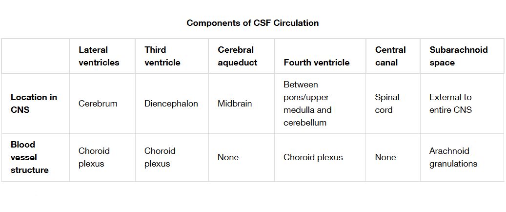
Lateral ventricle The lateral ventricle is a C-shaped cavity in each cerebral hemisphere, where the choroid plexus initiates CSF production. It connects to the third ventricle via the interventricular foramen, serving as the starting point for CSF flow.
Interventricular foramen The interventricular foramen, or foramen of Monro, links the lateral ventricles to the third ventricle, allowing CSF to move forward in a controlled manner. This narrow passage helps regulate fluid pressure between ventricular compartments.
Third ventricle The third ventricle is a slit-like cavity between the thalami, receiving CSF from the lateral ventricles through the interventricular foramina. It connects to the fourth ventricle via the cerebral aqueduct, facilitating further CSF distribution.
Cerebral aqueduct The cerebral aqueduct, also known as the aqueduct of Sylvius, is a narrow channel in the midbrain that transports CSF from the third ventricle to the fourth ventricle. Its small diameter makes it a potential site for flow obstruction, impacting circulation.
Fourth ventricle The fourth ventricle is a diamond-shaped space between the cerebellum and pons, receiving CSF from the cerebral aqueduct. It serves as a transition point where CSF exits into the subarachnoid space through specific apertures.
Median aperture The median aperture, or foramen of Magendie, is a midline opening in the fourth ventricle’s roof, allowing CSF to flow into the cisterna magna. This aperture ensures CSF reaches the subarachnoid space surrounding the brainstem and cerebellum.
Lateral apertures The lateral apertures, or foramina of Luschka, are two lateral openings in the fourth ventricle, enabling CSF to enter the subarachnoid space on both sides. These openings enhance the spread of CSF for broader protection and nutrient delivery.
Subarachnoid space The subarachnoid space is the area between the arachnoid and pia mater, filled with CSF that cushions the brain and spinal cord against mechanical stress. It extends throughout the cranial and spinal regions, supporting waste removal and fluid exchange.
Arachnoid granulations The arachnoid granulations are protrusions of the arachnoid mater into the dural sinuses, where CSF is reabsorbed into the venous system. These structures maintain intracranial pressure by filtering CSF back into the bloodstream at a rate of 0.3-0.4 ml per minute.
Superior sagittal sinus The superior sagittal sinus is a large dural sinus running along the midline of the skull, receiving reabsorbed CSF from the arachnoid granulations. It drains this fluid, along with deoxygenated blood, into the internal jugular vein, completing the circulation cycle.
Anatomy of CSF Circulation Components
The chart illustrates a well-organized system that governs CSF movement through the brain and spinal cord. This anatomical framework ensures effective fluid dynamics and neural protection.
- The choroid plexus, located in the lateral, third, and fourth ventricles, produces CSF through a combination of passive filtration and active secretion from blood plasma.
- The ventricular system, including the lateral, third, and fourth ventricles, provides a structured pathway, with narrow connections like the cerebral aqueduct controlling flow volume.
- The subarachnoid space acts as a reservoir, extending from the brain to the spinal cord, where CSF bathes neural tissues and facilitates nutrient exchange.
- Arachnoid granulations, embedded in the dural sinuses like the superior sagittal sinus, serve as the primary sites for CSF reabsorption into the venous circulation.
Physiological Role in Intracranial Health
CSF circulation plays a pivotal role in protecting the brain and regulating intracranial pressure. Its physiological functions are essential for maintaining a stable neural environment.
- The choroid plexus generates CSF at a rate of 0.3-0.4 ml/min, with a total daily production of 500 ml, though only 120-150 ml is present at any time due to constant turnover.
- CSF reduces the brain’s effective weight to approximately 50 grams within the skull, acting as a shock absorber against trauma or sudden movements.
- The subarachnoid space allows CSF to circulate nutrients and remove waste, supporting metabolic activity and preventing toxin buildup.
- Arachnoid granulations reabsorb CSF into the superior sagittal sinus, balancing production and drainage to keep intracranial pressure within the normal range of 7-15 mmHg.
Clinical Significance and Diagnostic Tools
The components of CSF circulation are clinically important due to their involvement in various neurological conditions. Advanced tools help assess and manage these dynamics.
- Hydrocephalus can result from blockages in the cerebral aqueduct or overproduction of CSF, leading to increased intracranial pressure that may require shunt placement.
- The interventricular foramen’s narrowness can contribute to non-communicating hydrocephalus if obstructed, necessitating surgical intervention.
- Magnetic resonance imaging (MRI) and computed tomography (CT) scans visualize CSF pathways, detecting abnormalities like dilated ventricles or impaired flow.
- Lumbar puncture measures CSF pressure, aiding in the diagnosis of conditions such as pseudotumor cerebri or meningitis-related complications.
CSF Flow Pathways and Regulation
The chart outlines a systematic pathway for CSF from production to reabsorption, highlighting the regulatory mechanisms involved. This process ensures consistent fluid balance and neural support.
- CSF flows from the lateral ventricles through the interventricular foramina into the third ventricle, driven by ciliary motion and pressure gradients.
- The cerebral aqueduct channels fluid into the fourth ventricle, where median and lateral apertures release it into the subarachnoid space.
- The subarachnoid space allows CSF to circulate around the brain and spinal cord, with arachnoid granulations facilitating reabsorption into the superior sagittal sinus.
- This cycle maintains a turnover every 4-6 hours, adapting to changes in production rates or absorption efficiency to stabilize intracranial conditions.
In conclusion, the components of CSF circulation, as depicted in this chart, form a sophisticated system that protects and nourishes the central nervous system while managing intracranial pressure. This detailed understanding of CSF dynamics supports the development of effective diagnostic and therapeutic approaches, enhancing overall neurological care.

