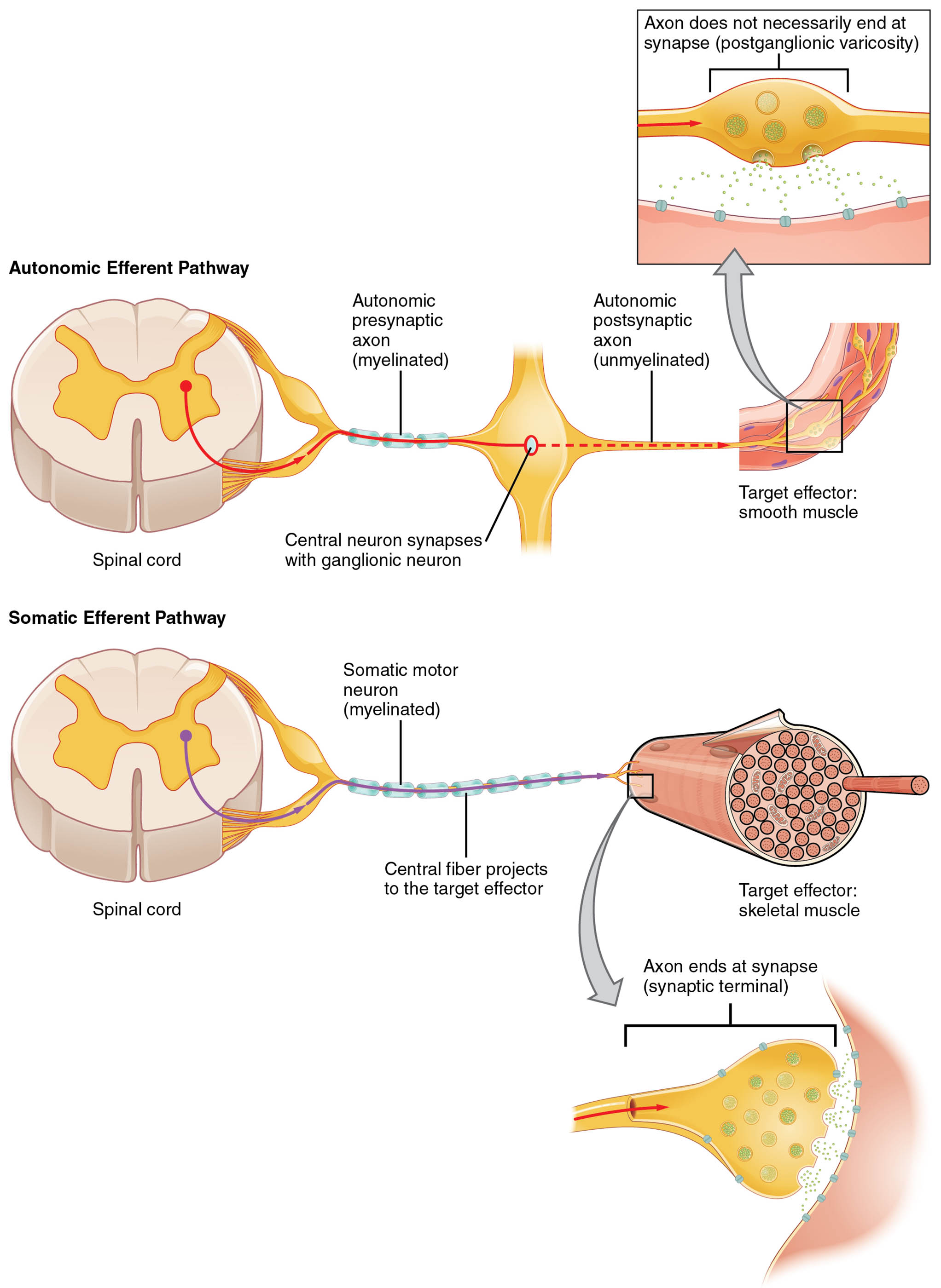
Reflexes are automatic responses that protect the body and maintain its functions, with somatic and visceral reflexes playing distinct yet complementary roles. This diagram highlights the similarities in afferent inputs and the differences in efferent pathways, showing how somatic reflexes directly connect to skeletal muscle via the ventral horn, while visceral reflexes involve a two-step process through ganglia to target effectors like smooth muscle or glands.
Labels in the Diagram
Afferent Neuron The Afferent Neuron carries sensory information from the periphery to the spinal cord, serving as the input for both somatic and visceral reflexes. It detects stimuli such as touch or pressure and relays them to the central nervous system.
Somatic Reflex Arc The Somatic Reflex Arc represents the pathway for voluntary muscle responses, originating from the afferent neuron. It involves a direct connection from the spinal cord’s ventral horn to skeletal muscle for rapid action.
Visceral Reflex Arc The Visceral Reflex Arc outlines the pathway for involuntary responses, also starting with the afferent neuron. It includes a projection to a ganglion and then to target effectors like the heart or digestive organs.
Central Neuron The Central Neuron is located in the spinal cord and processes sensory input from the afferent neuron. It initiates the efferent response in both reflex types, acting as the decision-making hub.
Ventral Horn The Ventral Horn is the region of the spinal cord’s gray matter containing motor neuron cell bodies for somatic reflexes. It sends direct signals to skeletal muscle via the efferent neuron.
Efferent Neuron The Efferent Neuron carries motor commands from the spinal cord to the target, differing between reflex types. In somatic reflexes, it directly innervates skeletal muscle, while in visceral reflexes, it involves a ganglion intermediary.
Skeletal Muscle The Skeletal Muscle is the target of the somatic reflex arc, responding to direct motor commands. It enables voluntary movements like withdrawing a hand from heat.
Ganglion The Ganglion is a cluster of neuron cell bodies in the visceral reflex pathway, receiving input from the central neuron. It then relays signals via a second efferent neuron to visceral effectors.
Second Efferent Neuron The Second Efferent Neuron extends from the ganglion to the target effector in visceral reflexes. It delivers the final signal to smooth muscle, cardiac muscle, or glands.
Smooth Muscle The Smooth Muscle is a visceral reflex target, found in organs like the stomach or blood vessels. It contracts or relaxes based on signals from the second efferent neuron.
Cardiac Muscle The Cardiac Muscle, located in the heart, is another visceral reflex target. It adjusts heart rate and force of contraction in response to autonomic input.
Glands The Glands, such as salivary or sweat glands, are visceral reflex effectors. They secrete substances like saliva or sweat following stimulation from the second efferent neuron.
Anatomy of Reflex Pathways
The somatic reflex arc and visceral reflex arc share a common starting point with the afferent neuron, which transmits sensory data to the central neuron in the spinal cord. However, their efferent branches diverge: the somatic reflex arc uses a single efferent neuron from the ventral horn to skeletal muscle, while the visceral reflex arc involves a ganglion and a second efferent neuron to reach smooth muscle, cardiac muscle, or glands.
- Spinal Cord Structure: The ventral horn houses alpha motor neurons for somatic control.
- Ganglion Role: Acts as an intermediary, containing preganglionic and postganglionic neurons.
- Effector Diversity: Includes voluntary skeletal muscle and involuntary visceral tissues.
This anatomical distinction reflects their specialized functions in the body.
Physiological Roles of Somatic and Visceral Reflexes
The somatic reflex arc enables quick, voluntary reactions to protect the body, such as the knee-jerk response. In contrast, the visceral reflex arc regulates involuntary processes, like adjusting heart rate via cardiac muscle or digestion via smooth muscle.
- Protective Response: The skeletal muscle pulls away from painful stimuli in somatic reflexes.
- Homeostatic Balance: The visceral reflex arc maintains organ function, such as glands secreting hormones.
- Speed of Action: Somatic reflexes are faster due to the direct efferent neuron path.
These reflexes work together to ensure both immediate safety and long-term stability.
Differences in Efferent Pathways
The somatic reflex arc features a straightforward connection from the ventral horn to skeletal muscle via the efferent neuron. The visceral reflex arc, however, requires a two-step process: the central neuron projects to a ganglion, followed by the second efferent neuron to smooth muscle or cardiac muscle.
- Single Synapse: Somatic reflexes use one synapse for efficiency.
- Two Synapses: Visceral reflexes involve a ganglion synapse for modulation.
- Neurotransmitter Use: Acetylcholine dominates somatic, while visceral may use acetylcholine or norepinephrine.
This structural difference supports their distinct physiological demands.
Clinical Relevance of Reflex Pathways
Understanding the somatic reflex arc and visceral reflex arc is vital for diagnosing neurological conditions. Damage to the ventral horn can impair skeletal muscle reflexes, while ganglion dysfunction may affect visceral reflex arc responses like digestion.
- Hyperreflexia: Overactive somatic reflex arc due to upper motor neuron lesions.
- Autonomic Dysreflexia: Disruption of the visceral reflex arc causing blood pressure spikes.
- Diagnostic Tests: Reflex hammers assess skeletal muscle response accuracy.
These insights guide effective treatment and rehabilitation strategies.
Evolutionary Perspective and Research Directions
The somatic reflex arc likely evolved for immediate survival, enabling quick skeletal muscle reactions to threats. The visceral reflex arc developed to support sustained organ function, with ganglion modulation enhancing adaptability.
- Evolutionary Benefit: Rapid efferent neuron responses protected against predators.
- Modern Studies: Explore ganglion plasticity in visceral reflex arc regulation.
- Therapeutic Advances: Target second efferent neuron pathways for autonomic disorders.
Ongoing research continues to uncover the complexities of these reflex systems.
In conclusion, the somatic reflex arc and visceral reflex arc demonstrate the body’s remarkable ability to respond to stimuli through distinct neural pathways. This diagram provides a clear comparison, from the afferent neuron to skeletal muscle or smooth muscle, offering valuable insights for anatomical study and clinical practice.

