Cerebrospinal fluid (CSF) is a vital component of the central nervous system, produced and circulated to cushion the brain and spinal cord while removing waste products. This article explores the pathway of CSF from its production in the choroid plexus through the ventricular system and subarachnoid space to its reabsorption into the bloodstream via the arachnoid granulations. Understanding this dynamic process offers insights into maintaining intracranial pressure and supporting overall neurological health.
Labeled Structures in CSF Circulation
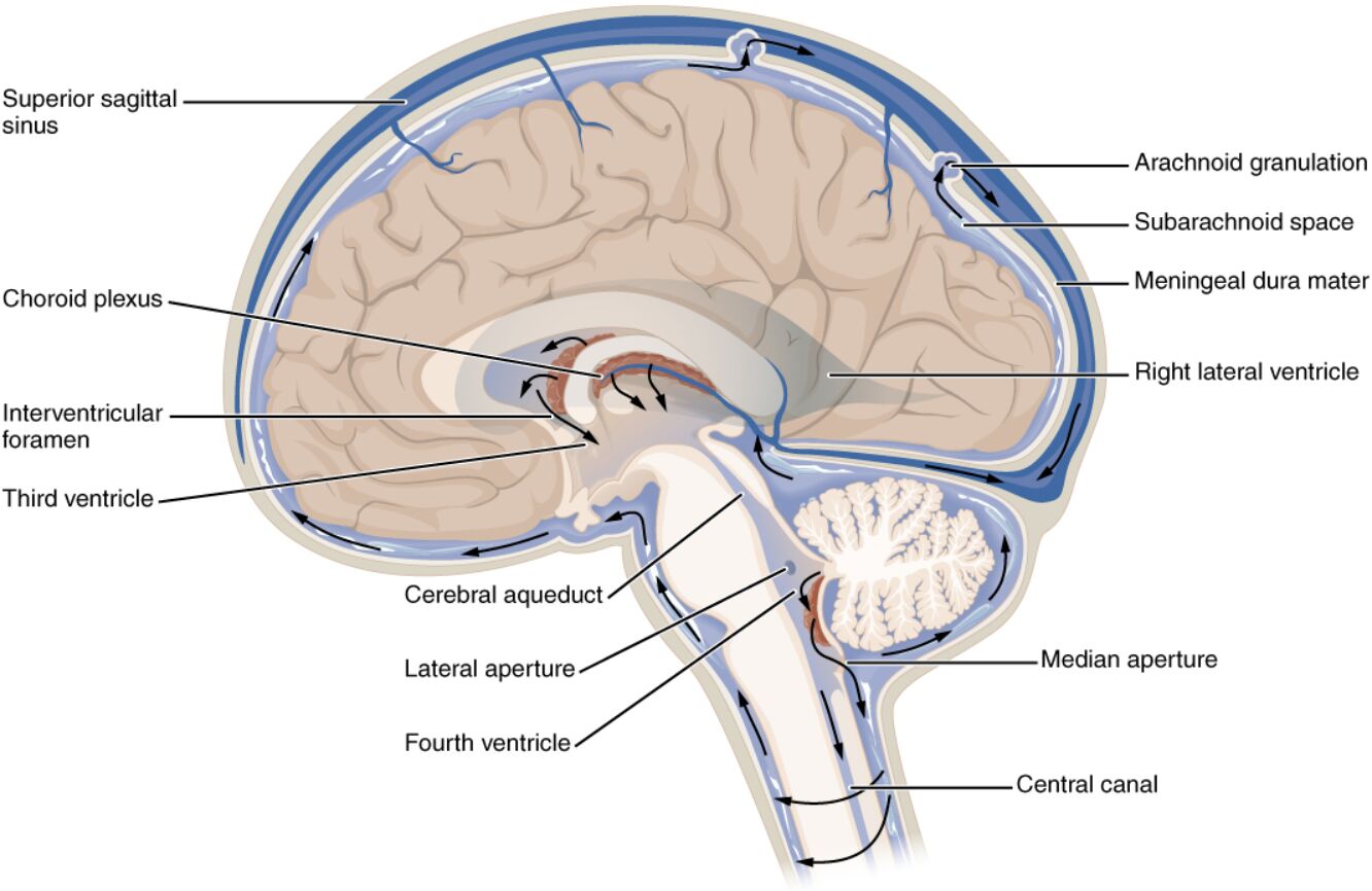
This section provides a detailed explanation of each labeled component in the provided medical image, highlighting their roles in CSF dynamics.
Choroid plexus The choroid plexus is a network of specialized ependymal cells located within the ventricles of the brain, responsible for producing approximately 500 ml of CSF daily. This structure secretes CSF into the ventricular system, maintaining a clear fluid that cushions and nourishes the brain and spinal cord.
Lateral ventricle The lateral ventricle is a paired C-shaped cavity within each cerebral hemisphere, where CSF is initially produced by the choroid plexus. It connects to the third ventricle via the interventricular foramen, allowing CSF to flow through the ventricular system.
Interventricular foramen The interventricular foramen, also known as the foramen of Monro, is a narrow passage linking the lateral ventricles to the third ventricle, facilitating CSF movement. It ensures a continuous flow of fluid, preventing pressure imbalances between ventricular compartments.
Third ventricle The third ventricle is a narrow, midline cavity between the thalami, receiving CSF from the lateral ventricles through the interventricular foramina. It connects to the fourth ventricle via the cerebral aqueduct, playing a key role in CSF distribution.
Cerebral aqueduct The cerebral aqueduct, or aqueduct of Sylvius, is a slender channel running through the midbrain, connecting the third and fourth ventricles. It allows CSF to pass from the third ventricle to the fourth, maintaining fluid equilibrium under normal conditions.
Fourth ventricle The fourth ventricle is a diamond-shaped cavity located between the cerebellum and pons, receiving CSF from the cerebral aqueduct. It serves as a conduit where CSF exits into the subarachnoid space through the median and lateral apertures.
Median aperture The median aperture, or foramen of Magendie, is a midline opening in the roof of the fourth ventricle, allowing CSF to flow into the cisterna magna of the subarachnoid space. This aperture ensures the distribution of CSF around the brainstem and cerebellum.
Lateral aperture The lateral aperture, or foramen of Luschka, consists of two lateral openings in the fourth ventricle, permitting CSF to enter the subarachnoid space on either side. These apertures enhance the spread of CSF, supporting its protective and nutritive roles.
Subarachnoid space The subarachnoid space is the area between the arachnoid mater and pia mater, filled with CSF that cushions the brain and spinal cord against trauma. It extends throughout the cranial and spinal compartments, facilitating waste removal and nutrient exchange.
Arachnoid granulations The arachnoid granulations are protrusions of the arachnoid mater into the dural sinuses, where CSF is reabsorbed into the venous bloodstream. These structures regulate intracranial pressure by filtering approximately 0.3-0.4 ml of CSF per minute into the superior sagittal sinus.
Dural sinus The dural sinus, such as the superior sagittal sinus, is a venous channel within the dura mater that receives CSF from the arachnoid granulations. It drains deoxygenated blood and reabsorbed CSF into the internal jugular vein, completing the circulation cycle.
Anatomy of CSF Circulation
The circulation of CSF involves a well-defined pathway that supports brain and spinal cord function. This anatomical layout ensures a steady supply and removal of fluid within the central nervous system.
- The choroid plexus, rich in capillaries and ependymal cells, produces CSF at a rate influenced by blood plasma filtration and active secretion.
- The ventricular system, comprising the lateral, third, and fourth ventricles, provides a conduit for CSF flow, with narrow passages like the cerebral aqueduct regulating volume.
- The subarachnoid space extends from the brain to the spinal cord, creating a reservoir that cushions neural tissue and allows CSF to bathe the surfaces.
- Arachnoid granulations, located along the dural sinuses, act as one-way valves, reabsorbing CSF into the venous system to maintain pressure homeostasis.
Physiological Role in Intracranial Dynamics
CSF circulation plays a critical role in protecting the brain and maintaining intracranial pressure. Its physiological functions are essential for neurological stability.
- The choroid plexus produces CSF at a rate of 0.3-0.4 ml/min, totaling about 500 ml daily, though only 120-150 ml is present at any given time due to constant turnover.
- CSF acts as a shock absorber, reducing the effective weight of the brain to about 50 grams within the skull, preventing compression against bony structures.
- The subarachnoid space facilitates nutrient delivery and waste removal, with CSF exchanging substances with the interstitial fluid of the brain.
- Arachnoid granulations reabsorb CSF into the dural sinuses, balancing production and drainage to keep intracranial pressure between 7-15 mmHg.
Clinical Significance and Imaging
The CSF circulation system is clinically significant due to its involvement in various neurological conditions. Advanced imaging techniques aid in assessing its function and detecting abnormalities.
- Hydrocephalus, caused by impaired CSF flow or absorption, can lead to increased intracranial pressure, treated with shunts or endoscopic third ventriculostomy.
- The cerebral aqueduct’s narrowness makes it a common site for obstruction, contributing to non-communicating hydrocephalus.
- Magnetic resonance imaging (MRI) and computed tomography (CT) cisternography visualize CSF pathways, identifying blockages or abnormal flow patterns.
- Monitoring CSF pressure via lumbar puncture helps diagnose conditions like pseudotumor cerebri, guiding therapeutic interventions.
CSF Flow Pathways and Regulation
The pathway of CSF from production to reabsorption involves a coordinated system that regulates fluid dynamics. This process ensures the brain’s protection and metabolic support.
- CSF flows from the lateral ventricles through the interventricular foramina into the third ventricle, driven by ciliary action and pressure gradients.
- The cerebral aqueduct channels fluid into the fourth ventricle, where median and lateral apertures release it into the subarachnoid space.
- The subarachnoid space allows CSF to circulate around the brain and spinal cord, with arachnoid granulations facilitating reabsorption into the dural sinuses.
- This cycle maintains a total CSF volume turnover every 4-6 hours, adapting to changes in intracranial pressure or metabolic demand.
In conclusion, the circulation of cerebrospinal fluid is a finely tuned process that protects and nourishes the central nervous system while managing intracranial pressure. The anatomical and physiological insights gained from studying this system enhance our ability to address neurological challenges, paving the way for improved diagnostic and therapeutic strategies.

