Introduction
This case presents a 5-year and 6-month-old male patient admitted with an acute asthma exacerbation, highlighting the clinical presentation, management, and relevant diagnostic findings. Understanding such cases is crucial for medical students and practitioners to develop proficiency in pediatric respiratory emergencies.
Case Presentation
The patient, a 5-year and 6-month-old male, presented to the emergency department yesterday morning with a chief complaint of cough. Upon triage, his oxygen saturation was measured at 87%, leading to immediate placement in the red zone. Evaluation in the red zone revealed widespread bilateral wheezing, prolonged expiration, and dyspnea. Oxygen support was initiated via a nebulizer mask, and the patient was transferred to the observation area where nebulized treatment commenced. Due to no significant improvement in his symptoms, the patient was admitted to the inpatient ward. This marks his first day of hospitalization for respiratory distress.
Past Medical History
The patient has no known diagnosis of asthma but has a history of frequent nebulizer use, starting at two years of age, occurring 7-8 times a year, with increased frequency during winter. This year alone, he has experienced 5-6 episodes. He has a nebulizer machine at home. The family reports a history of allergic rhinitis. He has two prior inpatient admissions for respiratory distress, lasting 14 days and 2-3 days, respectively. There is no history of food allergies during the complementary feeding period. The home environment is free of pets and dampness, but there is exposure to cigarette smoke.
Family History
His paternal aunt has a diagnosis of asthma.
Physical Examination (On Day 1 of Hospitalization)
- General Condition: Moderate, vitals stable.
- Skin: Normal, no rash.
- Head and Neck: No cervical lymphadenopathy.
- Oropharynx (OF): Normal.
- Respiratory System (SS): Respiratory distress, inspiratory and expiratory wheezing present (+), desaturates on room air.
- Cardiovascular System (CVS): S1+ S2+ (normal heart sounds), no extra sounds, no murmur heard, peripheral pulses palpable bilaterally.
- Gastrointestinal System (GIS): Abdomen soft, no tenderness, no rebound tenderness, bowel sounds present, gas and stool passage observed.
- Genitourinary System (GUS): Externally male, no hernia, no major urogenital anomalies, urine output present.
- Neuromuscular System (NMS): GCS: 15, conscious, pupils isochoric, light reflex +/+, neuromotor development appropriate for age, no meningeal irritation signs.
- Extremities: No deformities.
Chest X-ray Findings
The provided chest X-ray image (anterior-posterior view)
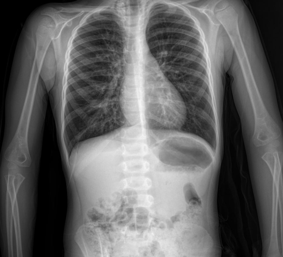
demonstrates several key features often seen in pediatric asthma exacerbations:
- Hyperinflation: While subtle, there are signs suggestive of hyperinflation, characterized by flattened diaphragms and increased retrosternal air space. This is due to air trapping in the lungs, a hallmark of bronchoconstriction and airway obstruction in asthma.
- Peribronchial Cuffing: There is evidence of peribronchial cuffing, appearing as increased density around the bronchi. This indicates bronchial wall thickening due to edema and inflammation, contributing to airway narrowing.
- Increased Bronchovascular Markings: The lung fields show increased prominence of bronchovascular markings, reflecting the inflammatory process and potentially some atelectasis or mucous plugging in smaller airways.
- No Focal Consolidation or Pneumothorax: Importantly, there is no evidence of focal consolidation, which would suggest pneumonia, or pneumothorax, a potentially life-threatening complication. This helps to narrow down the differential diagnosis in a child presenting with acute respiratory distress.
- Normal Cardiac Silhouette: The cardiac silhouette appears normal in size and contour, indicating no cardiomegaly or other significant cardiac pathology contributing to the respiratory symptoms.
Treatment
The patient is currently receiving the following treatment regimen:
- Salbutamol with proper doses
- Ipratropium Bromide with proper doses (likely referring to frequency and dose)
- Budesonide nebulized with proper doses
- Prednisolone IV with proper doses
- Magnesium Sulfate IV with proper doses
- Clarithromycin PO with proper doses
Discussion
This case exemplifies a typical presentation of an acute asthma exacerbation in a young child with a significant history of recurrent respiratory issues, despite lacking a formal asthma diagnosis prior to this admission. The presence of bilateral wheezing, prolonged expiration, and desaturation are classic signs. The chest X-ray findings, particularly hyperinflation and peribronchial cuffing, are consistent with an asthma flare-up, while ruling out other acute lung pathologies. The comprehensive treatment regimen, including bronchodilators, corticosteroids, magnesium sulfate, and antibiotics (likely for potential co-existing infection or as a broad-spectrum approach given the severity), reflects standard management protocols for severe asthma exacerbations. The family history of asthma and personal history of allergic rhinitis further support the underlying atopic predisposition common in asthmatic patients. Long-term management will likely involve formal asthma diagnosis, trigger avoidance strategies, and a tailored maintenance therapy plan.

