Introduction to Bone Imaging
Skeletal X-rays are fundamental for diagnosing a wide range of bone conditions, from traumatic injuries to metabolic and genetic disorders. This article presents a fascinating case of an adolescent girl initially investigated for severe hypertension and renal artery stenosis, where widespread sclerotic bone lesions were incidentally discovered, leading to the diagnosis of hereditary osteopoikilosis.
Clinical Presentation
A 15-year-old female patient presented to the emergency department on November 14, 2025, with complaints of nausea, abdominal pain, stomach cramps, and dizziness. Upon admission, her blood pressure was critically elevated at 185/110 mmHg, which later stabilized to 162/110 mmHg and 157/99 mmHg on subsequent measurements. Blood tests and an electrocardiogram (ECG showed normal sinus rhythm, QTc: 0.38) were performed, and she was admitted for further investigation and management of her hypertension.
Diagnostic Workup for Hypertension:
- Doppler Ultrasound (14.11.2025) Report:
- Left Renal Artery: Resistive Index (RI) measured at 0.62.
- Right Renal Artery: RI measured at 0.41, indicating a decrease.
- Finding: Parvus et tardus waveform pattern observed distally in the right renal arcuate arteries, suggestive of a proximal stenosis.
- CT Angiography Report (Renal Vessels):
- Right renal artery is duplicated (an anatomical variation).
- Finding: Approximately 60-63% stenosis observed in the dominant segment of the right renal artery.
Based on these findings, the patient was admitted for close monitoring and treatment due to suspected renal artery stenosis. Consultation with the pediatric nephrology department led to plans for a PET scan, Holter monitoring, an interventional radiology appointment at an external center, and continued inpatient care.
Physical Examination Findings (Initial Admission):
- General Condition: Moderate-good, vital signs stable.
- Skin: Natural, no rashes observed.
- Oropharynx: Hyperemic.
- Head and Neck: No cervical lymphadenopathy.
- Respiratory System: Clear breath sounds on auscultation (HİHTSEK), no rales, no rhonchi, eupneic, no intercostal retractions.
- Cardiovascular System: Normal heart sounds (S1, S2) with no additional sounds or murmurs. Peripheral pulses are palpable bilaterally.
- Gastrointestinal System: Abdomen is soft, non-tender, with no defense or rebound tenderness. Bowel sounds present, with normal gas and stool passage.
- Neuromuscular System: Pupils are isochoric and reactive to light. Neuromotor development is appropriate for age. No signs of meningeal irritation.
- Extremities: No deformities noted.
Hospital Course and Transfer:
On Day 1 of hospitalization for renal artery stenosis and hypertension, the 15-year and 1-month-old female patient was transferred to Çam and Sakura City Hospital Pediatric Nephrology Service for renal angiography and subsequent follow-up.
Findings from Çam and Sakura City Hospital (Repeat Imaging):
- Renal Artery Color Doppler USG Examination:
- Both kidneys are normal in size, localization, and contours, with normal parenchymal thickness and echogenicity.
- No pelvicalyceal ectasia observed bilaterally. No significant size difference between kidneys.
- No sonopathology observed in bilateral adrenal lodges.
- Left RI: 0.62.
- Right RI: 0.41 (decreased).
- Parvus et tardus waveform pattern observed distally in the right renal arcuate arteries.
- CT Angiography Renal Vessels:
- Right renal artery is duplicated (variation).
- Approximately 60-63% stenosis observed in the dominant segment of the right renal artery.
Physical Examination Findings (Upon Transfer):
- General Condition: Moderate-good, BP: 111/67 mmHg. Right upper arm blood pressure minimally elevated (KVAH minimal – likely referring to brachial-ankle pulse wave velocity or similar index).
- Skin: Natural, no rashes observed.
- Oropharynx: Hyperemic.
- Head and Neck: No cervical lymphadenopathy.
- Respiratory System: Clear breath sounds, no rales, no rhonchi, eupneic, no intercostal retractions.
- Cardiovascular System: Normal heart sounds (S1, S2) with no additional sounds or murmurs. Peripheral pulses are palpable bilaterally.
- Gastrointestinal System: Abdomen is soft, non-tender, with no defense or rebound tenderness. Bowel sounds present, with normal gas and stool passage.
- Genitourinary System: Female external genitalia, with adequate urine output.
- Neuromuscular System: Pupils are isochoric and reactive to light. Neuromotor development is appropriate for age. No signs of meningeal irritation.
- Extremities: No deformities noted.
Radiological Findings: Skeletal X-rays (Incidental Discovery)
During the extensive workup for her hypertension and renal issues, skeletal X-rays were performed, which revealed a striking and unexpected finding.
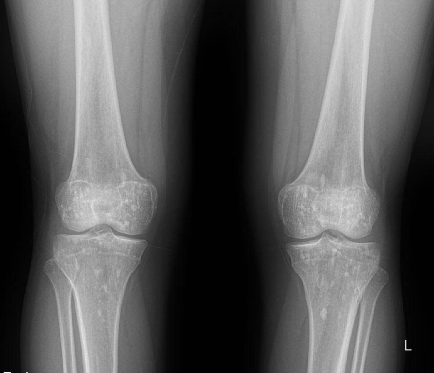
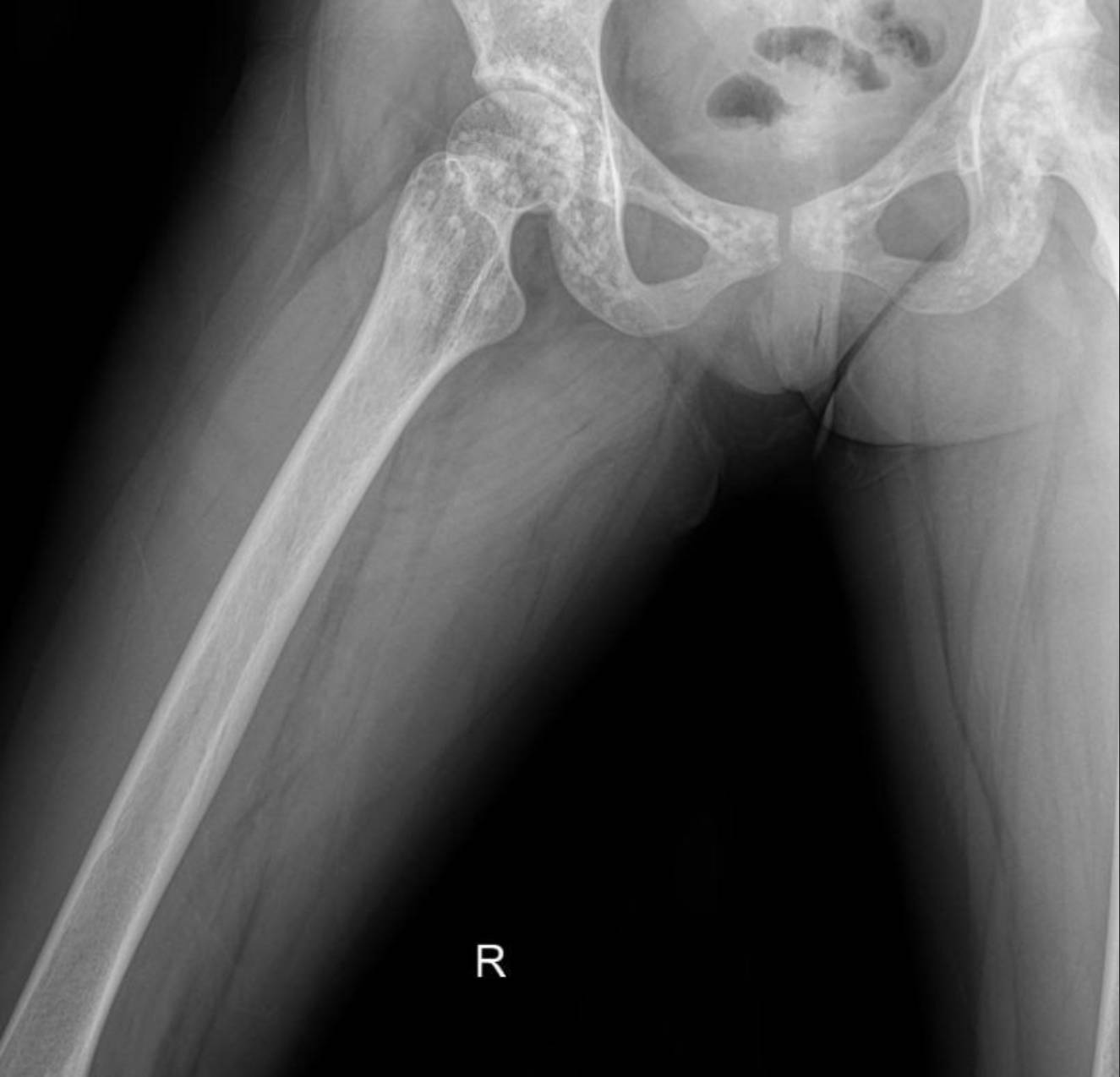
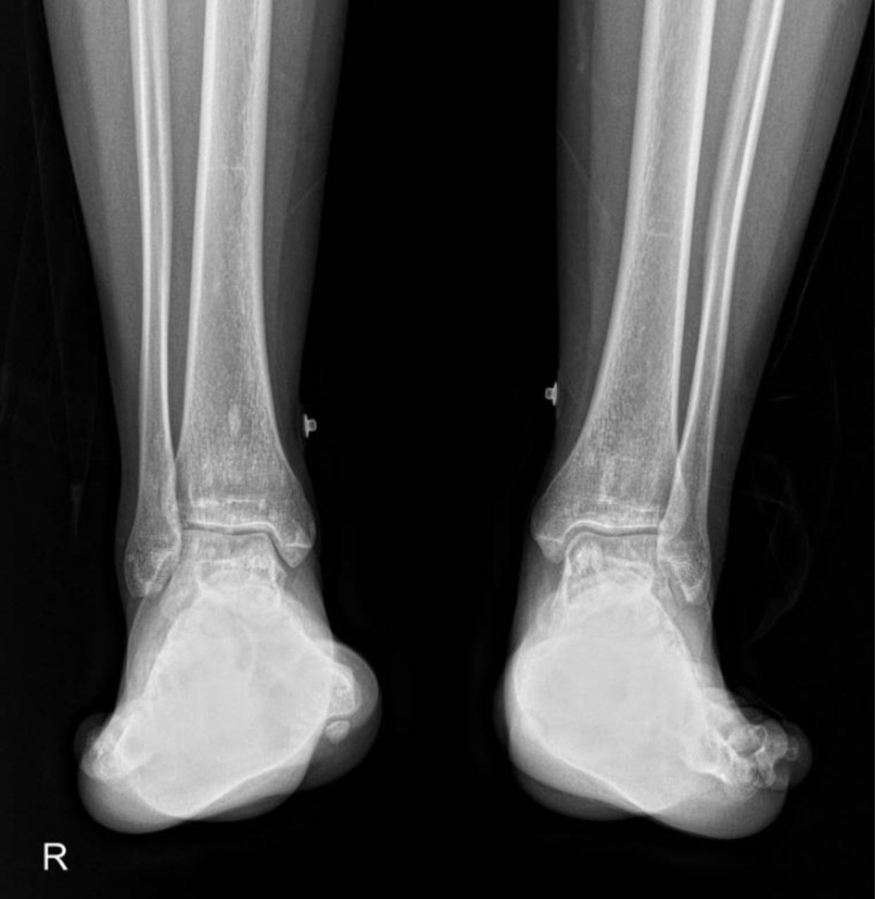
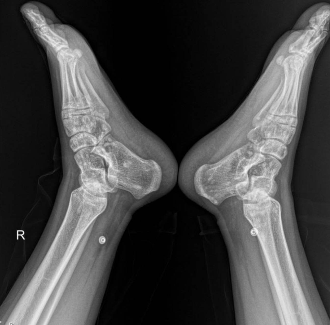
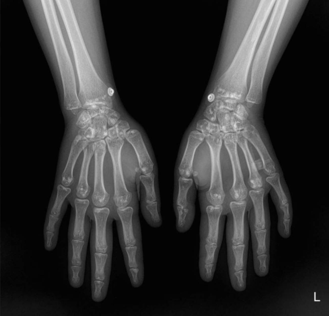
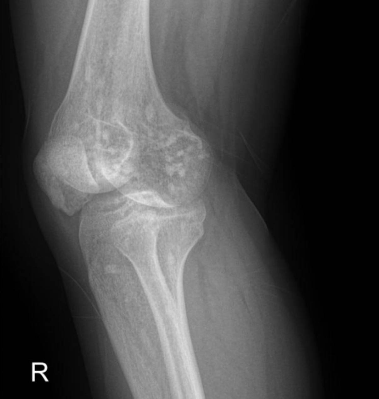

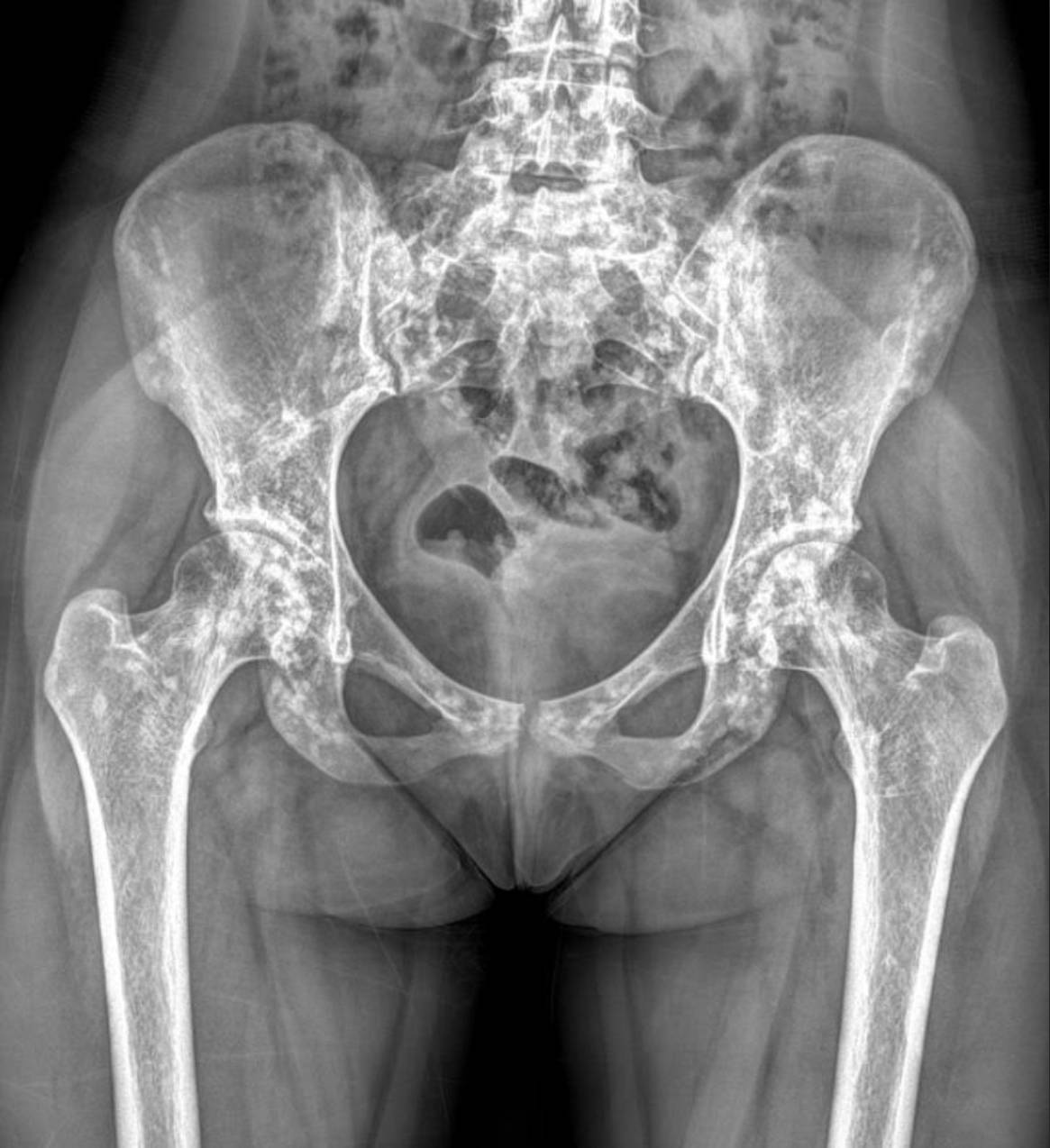
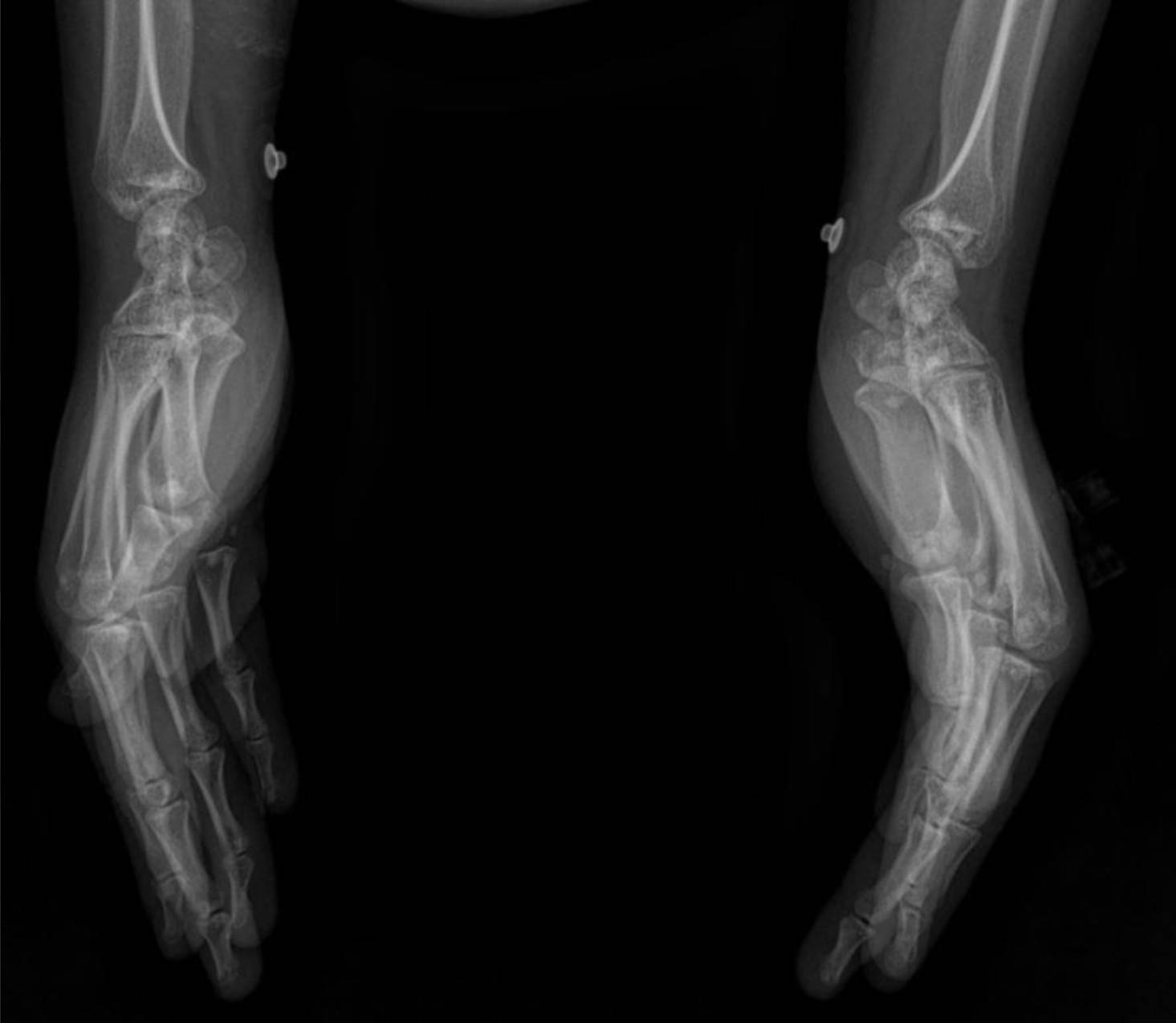
The X-rays of the pelvis, knees, elbows, wrists, and ankles demonstrate multiple, discrete, well-circumscribed sclerotic (densely opaque) lesions distributed symmetrically throughout the bones. These lesions are typically ovoid or spherical, varying in size, and are predominantly located in the epiphyses and metaphyses of long bones, as well as in the carpal and tarsal bones and the pelvic girdle.
Specifically:
- Pelvis and Hips: Numerous small, dense, rounded opacities are scattered throughout the iliac bones, sacrum, ischium, pubic rami, and around the femoral heads and greater trochanters.
- Knees: Both distal femurs and proximal tibiae show widespread small, dense sclerotic foci within the epiphyses and metaphyses. The joint spaces appear preserved.
- Elbows: Similar sclerotic lesions are evident in the distal humerus and proximal ulna and radius around both elbow joints.
- Wrists and Hands: The carpal bones, distal radius and ulna, and to a lesser extent, the metacarpals and phalanges, exhibit multiple small, dense foci.
- Ankles and Feet: The distal tibiae and fibulae, as well as the tarsal and metatarsal bones, show the characteristic sclerotic lesions.
These widespread, symmetrically distributed sclerotic bone islands are pathognomonic for hereditary osteopoikilosis (also known as osteopathia condensans disseminata). This condition is typically benign and often discovered incidentally. While not directly related to her renal artery stenosis or hypertension, its discovery is crucial for patient management as it can sometimes be associated with other connective tissue disorders, though often it is an isolated finding.
Conclusion
This case highlights a unique situation where a patient presenting with severe hypertension due to renal artery stenosis was incidentally diagnosed with hereditary osteopoikilosis through skeletal X-rays. While the two conditions are unrelated, the discovery of this rare genetic bone disorder is important for a complete patient profile. It underscores the value of thorough imaging in complex cases, sometimes revealing unexpected findings with significant clinical implications for patient counseling and long-term follow-up.
Please note: This content is intended for educational purposes and reference only. It should not be used as a substitute for professional medical advice, diagnosis, or treatment. Always seek the advice of a qualified healthcare provider for any medical concerns.

