Introduction to Pediatric Thoracic Imaging
Pediatric chest X-rays are invaluable in diagnosing respiratory illnesses in children. Accurate interpretation requires a thorough understanding of pediatric anatomy and common pathologies. This article presents a case study of a young girl with respiratory symptoms, focusing on the interpretation of her chest X-ray and the associated clinical context and management.
Clinical Presentation
The patient is a 2-year and 9-month-old female. She presented to the pediatric emergency department after experiencing a runny nose and cough for three weeks, with symptoms worsening and fever developing over the last four days. Upon arrival, she exhibited respiratory distress and an oxygen saturation (SpO2) of 91%. Nebulizer treatment and antibiotic therapy were initiated immediately. Due to desaturation in room air, the patient was admitted to the ward for continuous monitoring with a nebulizer mask and ongoing treatment.
Relevant History:
- No prior history of ward admission, intensive care unit (ICU) admission, or nebulizer treatment.
- Diagnosed with dust and pollen allergy 3 months prior, for which montelukast was prescribed.
- Family History: Paternal grandmother has asthma, father had suspected asthma as a child, and an aunt has a history of allergies.
- Environmental Factors: No smokers at home, no dampness or pets.
Physical Examination Findings:
- General Condition: Moderate, vital signs stable.
- Skin: Natural, no rashes observed.
- Head and Neck: Normal, no cervical lymphadenopathy.
- Oropharynx: Within normal limits.
- Respiratory System: Exhibits inspiratory and expiratory rhonchi and crackles (rales+), eupneic, with no intercostal retractions (İKÇ -), monitored with a nebulizer mask.
- Cardiovascular System: Normal heart sounds (S1, S2) with no additional sounds or murmurs.
- Gastrointestinal System: Abdomen is soft, non-tender, with no defense or rebound tenderness. Bowel sounds present, with normal gas and stool passage.
- Genitourinary System: No major urogenital anomalies externally observed, with adequate urine output.
- Neuromuscular System: Glasgow Coma Scale (GCS) of 15. Conscious, pupils are isochoric and reactive to light. No signs of meningeal irritation.
- Extremities: No deformities noted.
Radiological Findings: Chest X-ray
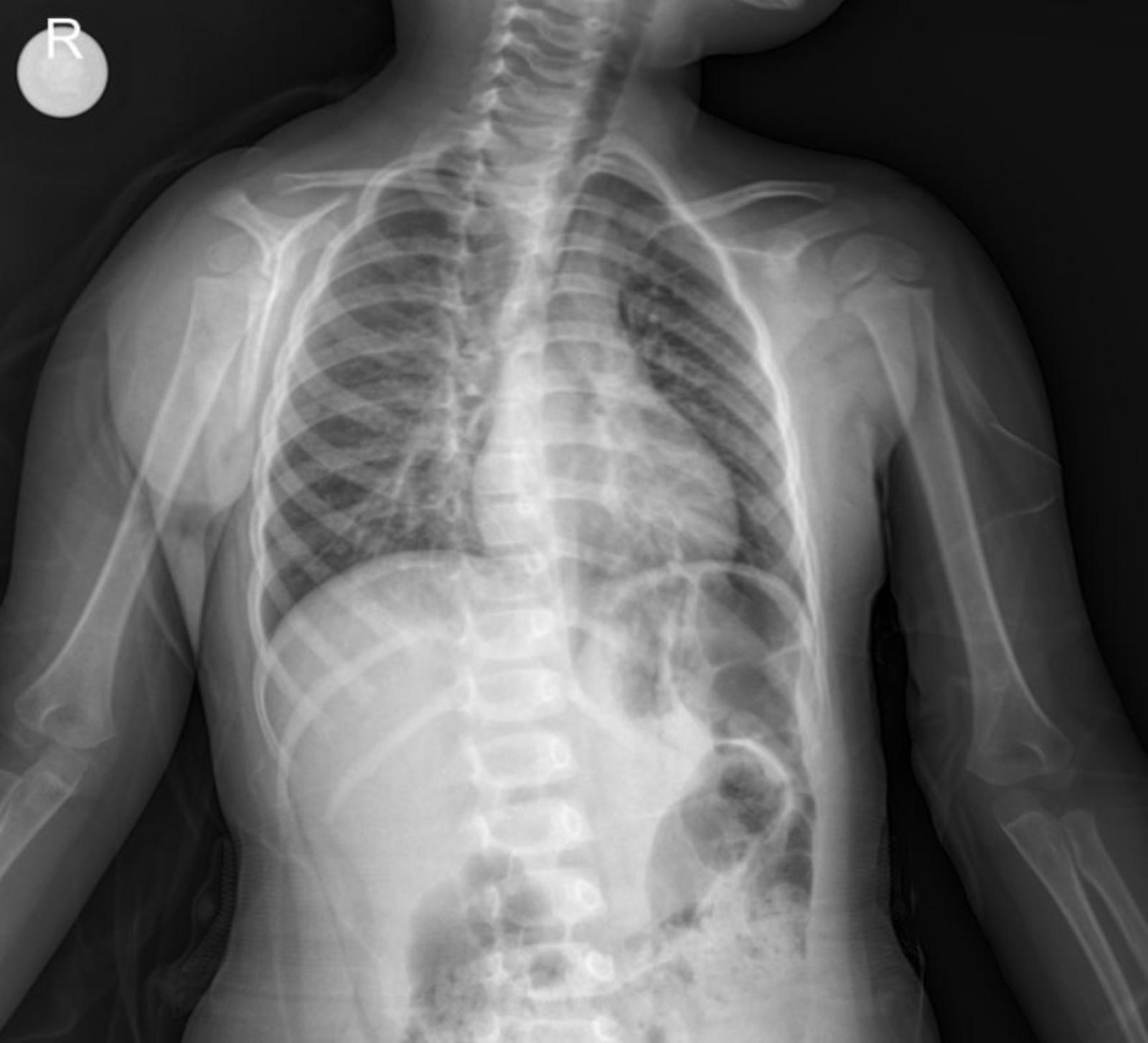
The provided anterior-posterior (AP) chest X-ray of the child reveals several significant findings:
- Lungs: The lung fields show evidence of peribronchial thickening and increased interstitial markings, consistent with an inflammatory process. Importantly, there is prominent paracardial infiltration, particularly noted around the left cardiac border. This suggests areas of consolidation or inflammation in the lung parenchyma adjacent to the heart, which is a common finding in bronchopneumonia.
- Heart: The cardiac silhouette appears within normal limits for size and contour for a pediatric patient, despite the adjacent infiltration.
- Diaphragm: The diaphragmatic contours are generally well-defined, with no evidence of significant pleural effusions.
- Bony Thorax: The ribs and clavicles appear intact, with no obvious fractures or deformities. The skeletal structures are appropriate for the child’s age.
- Trachea: The trachea appears patent and centrally located.
Considering the clinical history of worsening respiratory symptoms, fever, and hypoxemia, coupled with the X-ray findings of diffuse inflammatory changes and distinct paracardial infiltration, the diagnosis of bronchopneumonia is well supported.
Management Plan (Day 1 of Hospitalization for Bronchopneumonia)
The patient is currently receiving the following treatments:
- Antibiotics:
- Ampicillin/Sulbactam intravenous administration with proper doses.
- Azithromycin oral administration with proper doses.
- Bronchodilators:
- Salbutamol inhalation with proper doses.
- Ipratropium bromide inhalation with proper doses.
- Corticosteroids:
- Budesonide inhalation with proper doses.
- Methylprednisolone intravenous administration with proper doses.
- Other Medications:
- Magnesium sulfate intravenous administration with proper doses.
Disclaimer: Drug types and dosages should always be confirmed with the relevant dosage guidelines in your country or by consulting a licensed physician in the department. This information is for educational purposes only.
Conclusion
This case effectively demonstrates how a combination of detailed clinical history, physical examination findings, and radiological evidence from a chest X-ray leads to the diagnosis and appropriate management of pediatric bronchopneumonia with paracardial infiltration. The presence of paracardial infiltration on the X-ray further characterizes the extent of the inflammatory process.
Please note: This content is intended for educational purposes and reference only. It should not be used as a substitute for professional medical advice, diagnosis, or treatment. Always seek the advice of a qualified healthcare provider for any medical concerns.

