Bronchopulmonary dysplasia (BPD) X-ray image summary, anatomy, findings, differential diagnosis and treatment
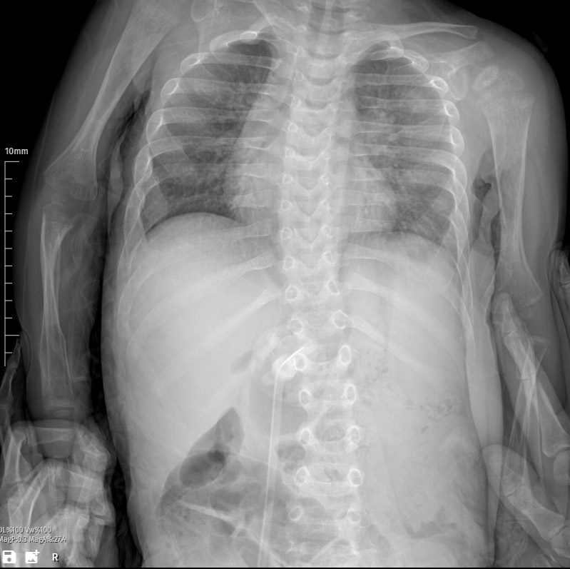
This chest X-ray depicts a 1-year-old with bronchopulmonary dysplasia (BPD), a chronic lung condition stemming from premature birth and prolonged mechanical ventilation. The radiograph serves as a pivotal diagnostic tool, offering insights into the extent and asymmetry of pulmonary infiltrates in this vulnerable population, crucial for tailored management strategies.
Clinical Summary:
In the intricate landscape of pediatric respiratory health, this patient’s history of BPD raises concerns about the intricate interplay of prematurity, prolonged ventilatory support, and the potential exacerbation of chronic lung disease. The X-ray serves as a pivotal diagnostic element, guiding clinical decisions to optimize respiratory support and address acute exacerbations.
Radiological Findings:
The X-ray reveals bilateral infiltrates, indicative of inflammatory or infectious processes affecting the lung parenchyma. Notably, the left lung exhibits more prominent infiltrative changes, highlighting an asymmetrical distribution of pathology. This observation prompts a closer examination of the localized exacerbation in the context of BPD.
Anatomical Localization:
Bilateral infiltrates extend across both lung fields, with a distinct emphasis on the left lung. This anatomical insight informs clinical reasoning, indicating a localized aggravation of the underlying pathology, potentially influencing therapeutic interventions and prognostic considerations for this pediatric patient.
Clinical Implications:
In the realm of BPD, infiltrates on the X-ray signify an acute exacerbation, predisposing the patient to compromised respiratory function, increased work of breathing, and heightened susceptibility to respiratory infections. The distinct asymmetry in lung involvement necessitates a nuanced approach to address the unique challenges posed by this exacerbation.
Differential Diagnosis:
Considering the vulnerability of children with BPD, a comprehensive differential diagnosis encompasses infectious etiologies such as viral or bacterial pneumonias. Non-infectious causes, including aspiration or exacerbation of the underlying chronic lung disease, should also be explored. A precise diagnosis is imperative for targeted therapeutic interventions.
Treatment Options:
Management involves a multi-pronged approach addressing the acute exacerbation and the underlying BPD. Oxygen therapy may be essential for maintaining adequate oxygenation, while empiric antibiotics are initiated to cover potential bacterial infections. Close monitoring is paramount to assess treatment response, ensuring adjustments are made promptly based on evolving clinical and radiological parameters.
Recommendations:
- Close Monitoring: Regular clinical and radiological assessments to gauge the patient’s response to treatment and identify any evolving complications.
- Optimization of Ventilatory Support: Ensure appropriate ventilatory support tailored to the specific needs of the patient, taking into account the underlying bronchopulmonary dysplasia.
- Nutritional Support: Adequate nutritional support to promote growth and development, considering the increased metabolic demands associated with respiratory distress.
- Infectious Disease Workup: Conduct targeted investigations, including respiratory cultures and viral panels, to identify the causative agent and guide antimicrobial therapy.
In summary, this chest X-ray in a 1-year-old with bronchopulmonary dysplasia reveals bilateral infiltrates, notably more pronounced in the left lung. The interpretation underscores the complexity of managing respiratory distress in pediatric patients with chronic lung diseases.

