The human brain’s ability to process and produce language is a remarkable feat, largely driven by specialized regions within the cerebral cortex. This diagram highlights Broca’s area and Wernicke’s area, two critical integration zones connected by deep white matter, which together enable the comprehension and articulation of speech. Exploring these areas provides valuable insights into the neural basis of communication, offering a foundation for understanding how language shapes our interactions and cognitive experiences.
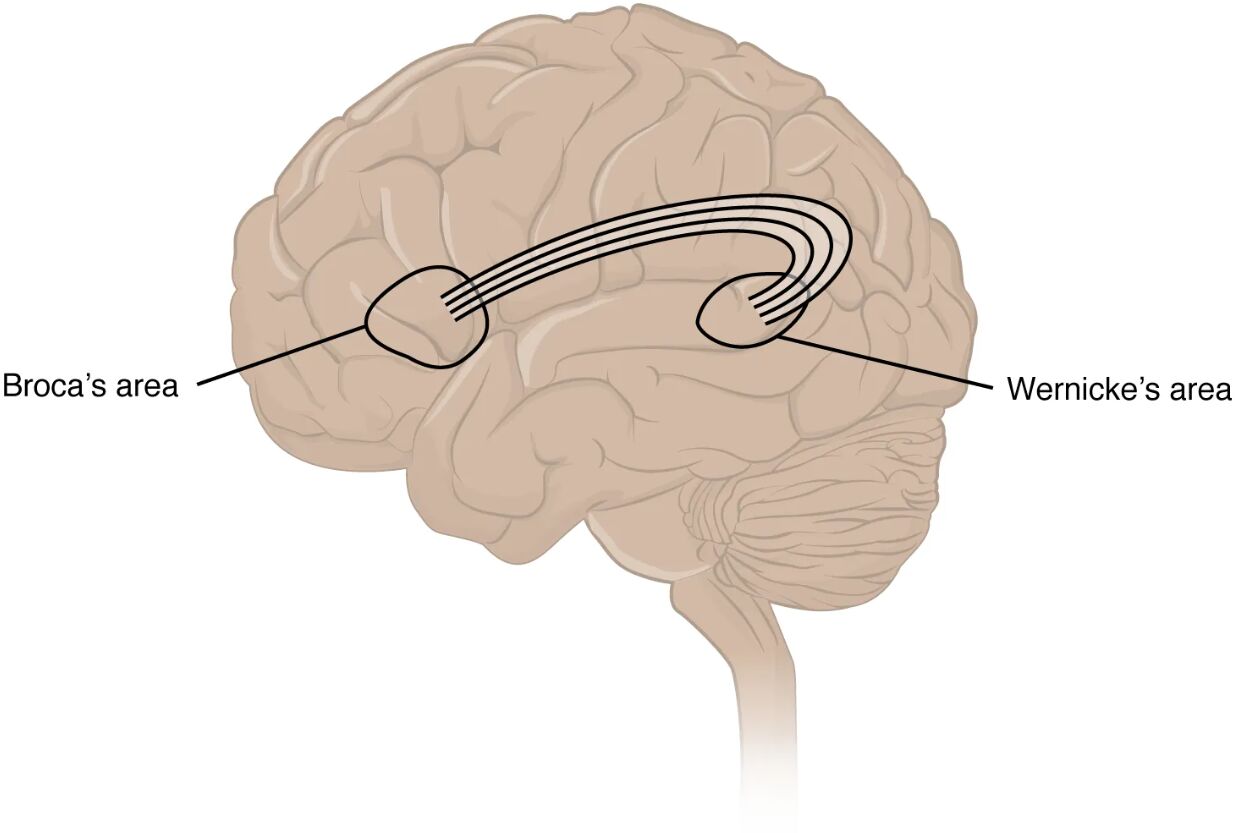
Broca’s area Broca’s area, located in the frontal lobe, is essential for speech production and the coordination of muscles involved in articulation. Damage to this region can result in expressive aphasia, where individuals struggle to form words despite understanding language.
Wernicke’s area Wernicke’s area, situated in the temporal lobe, plays a vital role in language comprehension and understanding spoken or written words. Impairment here can lead to receptive aphasia, characterized by difficulty in processing language meaning.
Anatomy of Language Processing Regions
The cerebral cortex houses specialized areas dedicated to language. This diagram illustrates their strategic locations and connections.
- Broca’s area resides in the left inferior frontal gyrus, typically dominant for language in most individuals.
- Wernicke’s area is located in the superior temporal gyrus, near the auditory cortex, facilitating sound-to-meaning conversion.
- These regions are linked by the arcuate fasciculus, a bundle of white matter fibers, enabling communication between them.
- The left hemisphere dominance for language is observed in about 95% of right-handed people and many left-handers.
- Blood supply from the middle cerebral artery supports these areas, making them susceptible to stroke-related damage.
Role of Broca’s Area in Speech Production
Broca’s area orchestrates the physical act of speaking. Its position in the frontal lobe enhances motor planning for language.
- This region controls the sequencing of muscle movements for speech, such as tongue and lip coordination.
- It integrates input from Wernicke’s area to translate thoughts into audible words.
- Damage can cause non-fluent speech, with effortful and halting articulation.
- Recovery often involves compensatory mechanisms in adjacent motor areas.
- Neuroimaging shows increased activity here during verbal tasks like naming objects.
Function of Wernicke’s Area in Language Comprehension
Wernicke’s area decodes the meaning behind sounds and words. Its proximity to auditory regions aids this process.
- This area processes phonological input, linking sounds to semantic understanding.
- It supports the ability to follow conversations or read with comprehension.
- Lesions here result in fluent but nonsensical speech, known as jargon aphasia.
- It connects to the angular gyrus, aiding in reading and writing integration.
- Functional MRI reveals activation during listening or language interpretation tasks.
The Arcuate Fasciculus: Connecting Language Centers
The arcuate fasciculus serves as the neural highway between Broca’s and Wernicke’s areas. This connection is crucial for cohesive language use.
- This white matter tract relays information from comprehension to production sites.
- It allows real-time feedback, enabling correction of speech errors during conversation.
- Disruption can lead to conduction aphasia, where repetition is impaired despite intact comprehension and production.
- The tract’s integrity is assessed using diffusion tensor imaging in neurological studies.
- Damage often results from trauma or demyelinating diseases affecting white matter.
Neurotransmitters and Hormonal Influences on Language
Chemical signaling enhances language function within these areas. These substances modulate neural activity and plasticity.
- Dopamine supports motivation and verbal fluency, influencing Broca’s area output.
- Acetylcholine facilitates memory retrieval, aiding Wernicke’s area comprehension.
- Serotonin regulates mood, indirectly affecting language-related emotional expression.
- Thyroid hormones like T3 and T4 boost metabolic activity, supporting cortical function.
- Imbalances can lead to language processing difficulties, necessitating further evaluation.
Clinical Implications of Language Area Damage
Understanding these regions aids in diagnosing and managing language disorders. This diagram supports clinical decision-making.
- Expressive aphasia from Broca’s area damage requires speech therapy focused on articulation.
- Receptive aphasia from Wernicke’s area injury needs comprehension exercises and support.
- Stroke is a common cause, with the left middle cerebral artery territory at risk.
- Imaging like CT or MRI localizes lesions, guiding rehabilitation strategies.
- Early intervention improves outcomes, leveraging neuroplasticity in undamaged areas.
Developmental Aspects of Language Areas
Language areas develop through childhood, shaped by experience and genetics. This diagram reflects their mature organization.
- Broca’s area matures with speech practice, strengthening motor pathways by age five.
- Wernicke’s area develops alongside auditory exposure, enhancing comprehension skills.
- Bilingualism increases connectivity in the arcuate fasciculus, boosting language flexibility.
- Critical periods in early life determine optimal language acquisition.
- Delays may indicate developmental disorders like specific language impairment.
Advances in Language Research
Ongoing studies deepen our understanding of these areas. This diagram inspires exploration into therapeutic innovations.
- Transcranial magnetic stimulation targets Broca’s area to enhance speech recovery.
- Functional connectivity studies map arcuate fasciculus integrity in real-time.
- Gene therapy explores repairing white matter damage affecting language tracts.
- Artificial intelligence models simulate language processing for training purposes.
- These advancements promise better outcomes for language rehabilitation.
In conclusion, this diagram of Broca’s and Wernicke’s areas provides a clear view of the cerebral cortex’s role in language, highlighting the intricate connection via the arcuate fasciculus. These regions work in tandem to enable the production and comprehension of speech, forming the basis of human communication. By exploring their anatomy and function, we gain insights into diagnosing and treating language disorders, paving the way for future research and improved patient care in the field of neuroscience.

