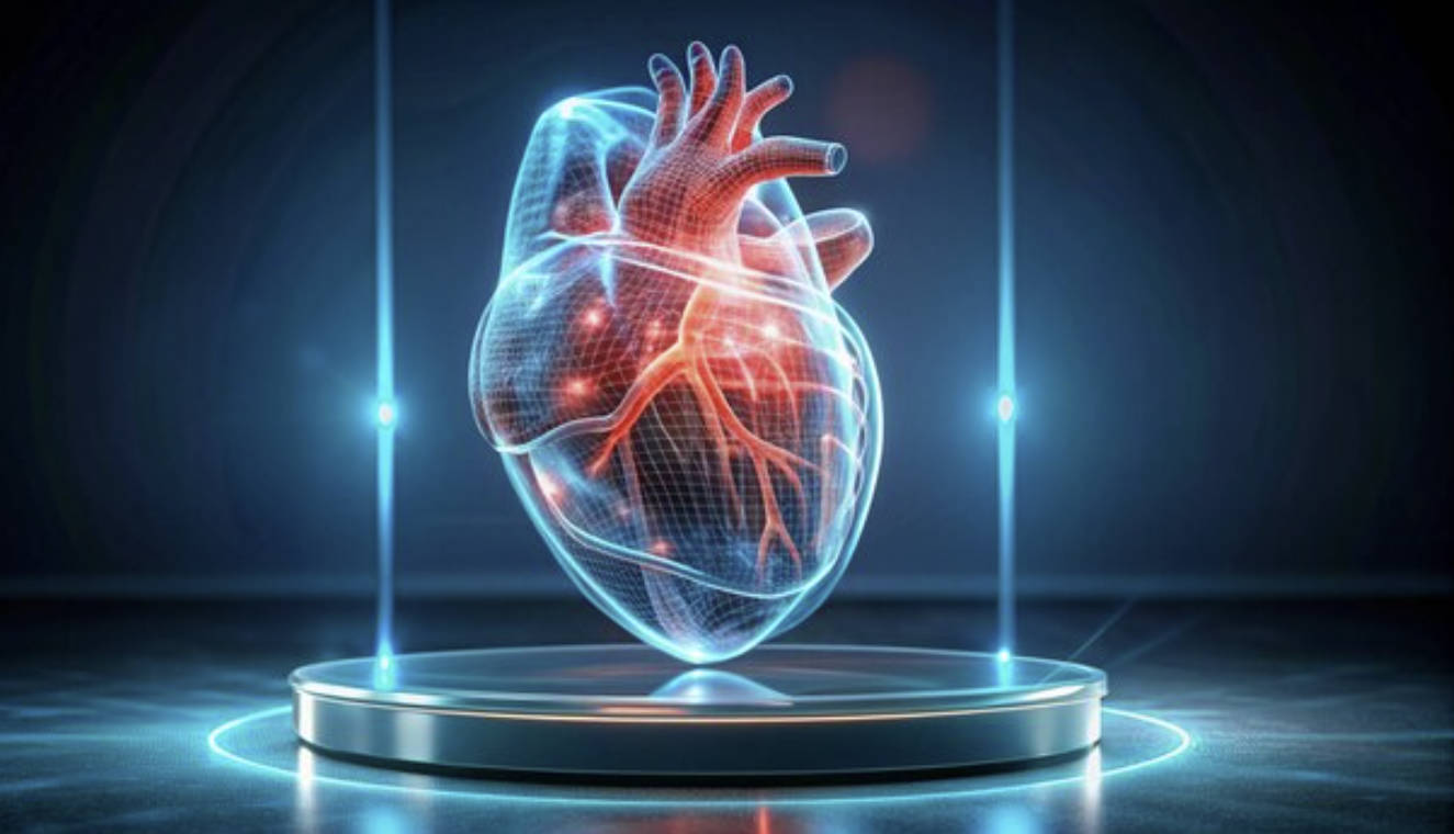
Scientists from the MRC Laboratory of Medical Sciences in the UK have created an innovative AI system called CardioKG that generates highly detailed views of heart structure and function. This tool has successfully uncovered previously unknown genes linked to heart diseases and even pointed to two existing drugs that could be repurposed for treatment. The development marks a significant step forward in speeding up gene discovery for diseases and improving the accuracy of predicting drug effectiveness, offering fresh hope for better heart disease management.
The research, led by postdoctoral researcher Dr. Khaled Rjoob and Professor Declan O’Regan, was published on December 28, 2025, in the journal Nature Cardiovascular Research.
The team built CardioKG by drawing on cardiac imaging data from the UK Biobank. They included scans from 4,280 patients diagnosed with atrial fibrillation, heart failure, or heart attacks, alongside data from 5,304 healthy individuals. By pulling out more than 200,000 image-derived features, they trained the model to capture a wide range of variations in how hearts look and work.
They then combined this imaging information with details pulled from 18 different biological databases. This integration allowed CardioKG to map out complex relationships and learn patterns in heart structure, function, genetics, and disease.
When put to the test, the system shone in several ways:
- It accurately predicted links between specific genes and cardiovascular diseases, spotting several genes that hadn’t been connected to heart conditions before.
- It highlighted opportunities for drug repurposing, suggesting that methotrexate (a medication typically used for rheumatoid arthritis) might help improve outcomes in heart failure.
- It also indicated that gliptins (a class of diabetes drugs) could offer benefits for patients with atrial fibrillation.
- Interestingly, the analysis revealed that caffeine may boost heart excitability and provide some protective effects for those with rapid, irregular heartbeats in atrial fibrillation.
The researchers believe CardioKG has huge potential beyond just cardiology. The same approach could be adapted for other organs, like the brain or body fat distribution, opening doors to new treatments for conditions such as dementia or obesity.
This kind of imaging-enhanced AI knowledge graph represents a powerful new tool in precision medicine, helping bridge the gap between detailed patient scans and broader biological knowledge to drive faster, more targeted discoveries.

