The human heart is a remarkable organ, tirelessly pumping blood to sustain life through a complex circulatory system. This diagram illustrates the key structures involved in blood circulation, offering a clear view of how oxygen-rich and oxygen-poor blood flows between the heart and lungs. Understanding these components is essential for grasping the fundamentals of cardiovascular anatomy and physiology.
Labelled Parts Explanation
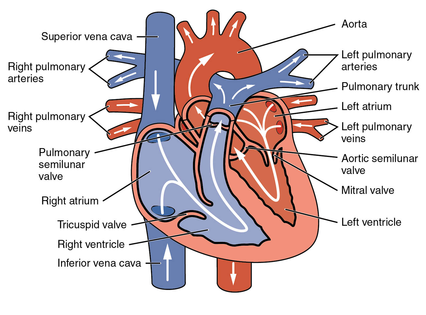
- Superior vena cava The superior vena cava is a large vein that carries deoxygenated blood from the upper body to the right atrium. It plays a critical role in returning blood to the heart for oxygenation.
- Right pulmonary arteries The right pulmonary arteries transport deoxygenated blood from the right ventricle to the lungs for oxygenation. These vessels branch out to ensure efficient gas exchange within the pulmonary circulation.
- Right pulmonary veins The right pulmonary veins return oxygenated blood from the lungs to the left atrium of the heart. This blood is then ready to be pumped into the systemic circulation.
- Pulmonary semilunar valve The pulmonary semilunar valve prevents backflow of blood from the pulmonary artery into the right ventricle during diastole. It ensures unidirectional flow as blood moves toward the lungs.
- Right atrium The right atrium receives deoxygenated blood from the body via the superior and inferior vena cava. It contracts to push blood into the right ventricle.
- Tricuspid valve The tricuspid valve separates the right atrium from the right ventricle, allowing blood to flow in one direction. It closes during ventricular contraction to prevent backflow into the atrium.
- Right ventricle The right ventricle pumps deoxygenated blood into the pulmonary arteries for oxygenation in the lungs. Its muscular walls are less thick compared to the left ventricle due to lower pressure requirements.
- Inferior vena cava The inferior vena cava brings deoxygenated blood from the lower body to the right atrium. It complements the superior vena cava in the venous return process.
- Aorta The aorta is the largest artery, carrying oxygenated blood from the left ventricle to the rest of the body. It distributes blood through its extensive network of branches.
- Left pulmonary arteries The left pulmonary arteries deliver deoxygenated blood from the right ventricle to the left lung for oxygenation. They are part of the pulmonary circulation system.
- Pulmonary trunk The pulmonary trunk is the main artery that carries deoxygenated blood from the right ventricle to the lungs. It divides into the left and right pulmonary arteries.
- Left atrium The left atrium receives oxygenated blood from the pulmonary veins. It then contracts to send blood into the left ventricle.
- Left pulmonary veins The left pulmonary veins transport oxygenated blood from the lungs to the left atrium. This blood is essential for systemic circulation.
- Aortic semilunar valve The aortic semilunar valve prevents backflow of blood from the aorta into the left ventricle. It opens during ventricular systole to allow blood ejection.
- Mitral valve The mitral valve, also known as the bicuspid valve, separates the left atrium from the left ventricle. It ensures blood flows unidirectionally into the ventricle.
- Left ventricle The left ventricle pumps oxygenated blood into the aorta for distribution to the body. Its thick, muscular walls generate the high pressure needed for systemic circulation.
Anatomical Overview of the Heart
The heart is a muscular organ located slightly to the left of the chest, serving as the central pump of the circulatory system. This diagram provides a detailed view of the heart’s internal structure, highlighting the flow of blood through its four chambers. The color coding—blue for deoxygenated blood and red for oxygenated blood—helps visualize the dual circulation process.
- The heart’s right side handles deoxygenated blood, receiving it from the body and sending it to the lungs.
- The left side manages oxygenated blood, distributing it to the body’s tissues via the aorta.
- Valves such as the tricuspid and mitral ensure efficient blood flow by preventing backflow.
- The pulmonary and aortic semilunar valves play a key role in maintaining pressure gradients during the cardiac cycle.
This anatomical layout is crucial for understanding how the heart maintains circulation, supporting oxygen delivery and waste removal.
The Role of Valves in Blood Circulation
Valves are essential components that regulate blood flow within the heart. They open and close in response to pressure changes during the cardiac cycle, ensuring efficient pumping.
- The tricuspid valve and mitral valve are atrioventricular valves that separate the atria from the ventricles.
- The pulmonary semilunar valve and aortic semilunar valve guard the exits of the right and left ventricles, respectively.
- These valves prevent regurgitation, maintaining the one-way flow critical for circulation.
- Dysfunction in these valves, such as stenosis or regurgitation, can lead to conditions like heart failure.
Their precise coordination ensures that blood moves smoothly from the atria to the ventricles and out to the pulmonary and systemic circuits.
Pulmonary and Systemic Circulation
The heart facilitates two distinct circulation paths: pulmonary and systemic. This dual system ensures efficient oxygenation and nutrient delivery.
- Pulmonary circulation begins with the right ventricle pumping blood into the pulmonary trunk, which splits into the right pulmonary arteries and left pulmonary arteries.
- In the lungs, blood is oxygenated and returns via the right pulmonary veins and left pulmonary veins to the left atrium.
- Systemic circulation starts with the left ventricle pushing blood into the aorta, distributing it to the body.
- The superior vena cava and inferior vena cava return deoxygenated blood to the right atrium, completing the cycle.
This process highlights the heart’s role in maintaining homeostasis by balancing oxygen and carbon dioxide levels.
Physiological Significance of Heart Chambers
The heart’s chambers work in harmony to sustain life. Each chamber has a specific function tailored to its anatomical position.
- The right atrium and left atrium act as receiving chambers, collecting blood from the veins.
- The right ventricle and left ventricle serve as pumping chambers, ejecting blood into the pulmonary and systemic arteries.
- The left ventricle’s thicker walls reflect its role in generating higher pressure for systemic circulation.
- The right ventricle’s thinner walls are adapted for the lower pressure of pulmonary circulation.
This structural difference ensures the heart efficiently meets the body’s diverse circulatory demands.
Conclusion
The diagram of blood circulation in the human heart offers a window into the intricate workings of this vital organ. By understanding the roles of the atria, ventricles, valves, and major vessels, one can appreciate how the heart sustains life through continuous pumping. This knowledge serves as a foundation for exploring cardiovascular health and potential interventions, making it an invaluable resource for anyone interested in human anatomy and physiology. Regular study of such diagrams enhances comprehension of the heart’s dynamic processes, encouraging a deeper interest in its remarkable functionality.


it’s just saying hi for the one who is working for this material is so good
Thanks for your supporting! have a nice day!