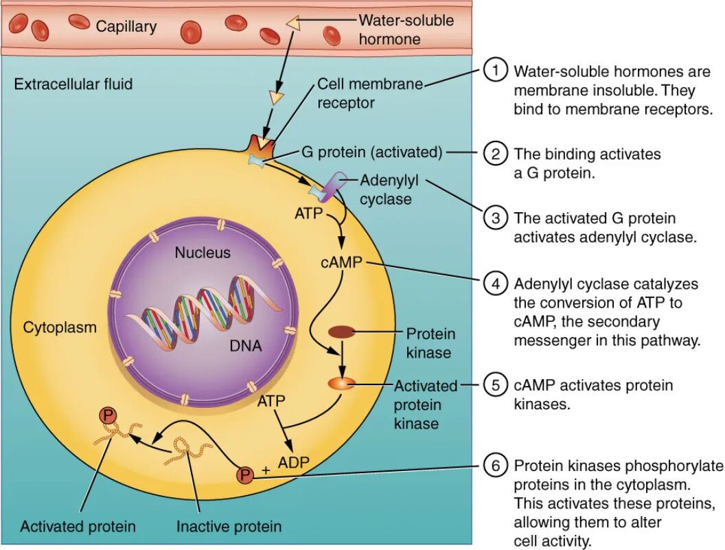
Water-soluble hormones are key regulators of cellular activity, unable to penetrate the cell membrane, which necessitates a unique signaling pathway within target cells. This diagram illustrates the process where a water-soluble hormone binds to a surface cell-membrane receptor, triggering a cascade involving G proteins, adenylyl cyclase, cyclic AMP (cAMP), and protein kinases, ultimately leading to the phosphorylation of proteins in the cytoplasm. Exploring this image offers a deeper understanding of how these hormones exert their effects through intricate intracellular signaling.
Labelled Parts Explanation
- Water-soluble hormone The water-soluble hormone, such as insulin or adrenaline, is a polar molecule that cannot diffuse through the lipid bilayer of the cell membrane. It initiates cellular responses by binding to specific receptors on the cell surface, triggering a signaling cascade.
- Surface cell-membrane receptor The surface cell-membrane receptor is a protein embedded in the plasma membrane that binds the water-soluble hormone with high affinity. This binding activates intracellular signaling pathways, such as those involving G proteins, to relay the hormone’s message.
- G proteins The G proteins are molecular switches located on the inner cell membrane that are activated upon receptor binding, transmitting the signal by interacting with effector enzymes. They cycle between active and inactive states, regulated by GTP and GDP, to amplify the hormonal response.
- Adenylyl cyclase The adenylyl cyclase is an enzyme activated by G proteins that converts ATP into cyclic AMP (cAMP), a secondary messenger. This activation step amplifies the signal, initiating a cascade that influences cellular functions like metabolism or secretion.
- Cyclic AMP (cAMP) The cyclic AMP (cAMP) is a second messenger produced by adenylyl cyclase that relays the signal within the cell, activating protein kinases. It plays a central role in amplifying and diversifying the hormone’s effects on target processes.
- Protein kinases The protein kinases are enzymes activated by cAMP that phosphorylate target proteins by adding phosphate groups, altering their activity. This phosphorylation triggers specific cellular responses, such as enzyme activation or gene expression changes.
- Phosphorylation of proteins The phosphorylation of proteins involves the addition of phosphate groups by protein kinases, activating or inhibiting these proteins to carry out the hormone’s intended effects. This step finalizes the signaling pathway, leading to physiological changes in the cell.
- Cytoplasm The cytoplasm is the cellular compartment where the signaling cascade, including protein phosphorylation, takes place, containing the machinery for these processes. It serves as the site where the hormone’s effects are translated into action within the cell.
Anatomical Overview of Water-Soluble Hormone Action
Water-soluble hormones, including peptides and catecholamines, exert their effects through a membrane-bound receptor mechanism rather than direct cellular entry. This diagram outlines the step-by-step signaling pathway that translates the hormone’s presence into cellular responses.
- The water-soluble hormone binds externally to the surface cell-membrane receptor.
- The G proteins and adenylyl cyclase initiate the second messenger system.
- The cyclic AMP (cAMP) and protein kinases amplify and execute the signal.
- The phosphorylation of proteins in the cytoplasm completes the process.
This pathway highlights the hormone’s indirect influence on cell function.
Role of the Water-Soluble Hormone
The water-soluble hormone serves as the initial trigger for cellular responses. Its binding is the critical first step.
- The water-soluble hormone includes insulin, produced by the pancreas, and adrenaline from the adrenal medulla.
- Its inability to cross the membrane necessitates surface receptor interaction.
- This binding specificity ensures targeted cellular effects.
- Examples regulate glucose levels or stress responses.
This step initiates the signaling cascade.
Function of the Surface Cell-Membrane Receptor
The surface cell-membrane receptor is the gateway for hormone action. Its activation drives intracellular events.
- The surface cell-membrane receptor binds the water-soluble hormone with high affinity.
- This binding activates associated G proteins to start the signaling pathway.
- The receptor’s location on the membrane allows external signal detection.
- Different receptors respond to specific hormones.
This interaction is essential for signal transmission.
Significance of G Proteins and Adenylyl Cyclase
G proteins and adenylyl cyclase amplify the hormonal signal. Their role is pivotal in the cascade.
- The G proteins switch to an active state, stimulating adenylyl cyclase.
- The adenylyl cyclase converts ATP to cyclic AMP (cAMP), boosting the signal.
- This amplification enhances the hormone’s reach within the cell.
- The process is tightly regulated to prevent overstimulation.
This step ensures effective signal propagation.
Process of cAMP and Protein Kinases
cAMP and protein kinases execute the signaling within the cell. Their activation drives physiological changes.
- The cyclic AMP (cAMP) activates protein kinases by binding to their regulatory subunits.
- The protein kinases phosphorylate target proteins, altering their function.
- This cascade amplifies the initial hormone signal.
- The effects can include enzyme activation or ion channel modulation.
This phase translates the signal into action.
Importance of Phosphorylation and Cytoplasm
Phosphorylation and the cytoplasm finalize the hormone’s impact. Their role completes the response.
- The phosphorylation of proteins activates or inhibits cellular proteins.
- The cytoplasm houses the ribosomes and enzymes for this process.
- This step produces the hormone’s intended physiological outcome.
- The cytoplasm integrates the signal with cellular metabolism.
This conclusion supports homeostatic regulation.
Physiological Importance of Water-Soluble Hormone Binding
The binding mechanism of water-soluble hormones ensures precise cellular regulation. Its efficiency supports homeostasis.
- The water-soluble hormone’s surface binding avoids direct membrane penetration.
- The G proteins, adenylyl cyclase, and cyclic AMP (cAMP) amplify the signal.
- The protein kinases and phosphorylation of proteins execute specific responses.
- This pathway adapts to rapid physiological demands.
The process is vital for metabolic and stress responses.
Clinical Relevance of Hormone Signaling
Understanding water-soluble hormone binding aids in diagnosing endocrine disorders. These components are key clinical markers.
- Overactive protein kinases can lead to uncontrolled cell growth, as in some cancers.
- Deficiency in cyclic AMP (cAMP) production may cause hormonal resistance.
- Abnormal G proteins function is linked to diseases like pseudohypoparathyroidism.
- Blood tests and receptor studies guide treatment.
This knowledge supports effective endocrine management.
Conclusion
The binding of water-soluble hormones diagram provides a detailed view of the process involving the water-soluble hormone, surface cell-membrane receptor, G proteins, adenylyl cyclase, cyclic AMP (cAMP), protein kinases, phosphorylation of proteins, and cytoplasm, illustrating how these hormones trigger intracellular signaling. By exploring the mechanism of receptor activation, second messenger production, and protein modification, one gains insight into the intricate cellular responses driven by these hormones. This understanding serves as a foundation for studying endocrinology and addressing related health concerns, encouraging further exploration of the sophisticated signaling pathways that regulate bodily functions.

