Balantidium coli is the only ciliated protozoan known to cause disease in humans, responsible for balantidiasis, a zoonotic intestinal infection that can range from asymptomatic carriage to severe dysentery and even life-threatening complications. This large ciliate, visible in the microscopic image, belongs to the supergroup Chromalveolata and possesses distinctive morphological features that allow it to thrive in the human large intestine. Understanding the structure and biology of Balantidium coli is essential for clinical microbiologists, infectious disease specialists, and public health professionals managing cases in regions with poor sanitation or close contact with pigs, the primary animal reservoir.
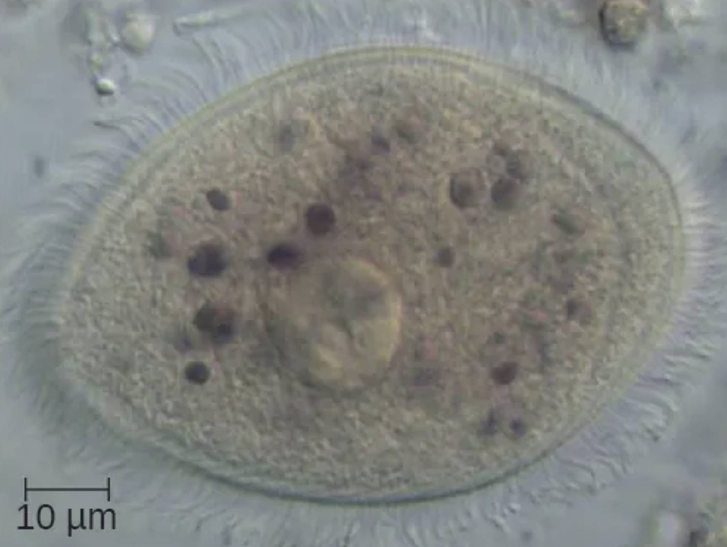
Balantidium coli is the large, oval-shaped ciliated protozoan shown in the image. Measuring approximately 50-100 μm in length, it is one of the largest protozoan parasites affecting humans and is characterized by its ovoid body covered in rows of cilia that enable rapid motility in the intestinal environment.
10 μm scale bar indicates the microscopic scale of the specimen, confirming the substantial size of this ciliate compared to other intestinal protozoa such as Entamoeba or Giardia. The scale helps clinicians and laboratory technicians accurately identify the organism during stool microscopy examinations.
Cilia are the numerous fine hair-like projections visible around the periphery of the organism. These structures beat in coordinated waves to propel the parasite through the intestinal lumen and facilitate feeding by creating water currents that bring bacteria and debris toward the cytostome, the ciliate’s mouth-like structure.
Macronucleus is the prominent, kidney-shaped or elongated large nucleus visible in the central region of the cell. It controls vegetative functions and is highly polyploid, supporting the high metabolic activity required for the parasite’s active lifestyle in the gut.
Micronucleus refers to the smaller, often less conspicuous nucleus located near the macronucleus. It is diploid and primarily involved in sexual reproduction through conjugation, although conjugation is rarely observed in clinical specimens.
Biology and Morphology of Balantidium coli
Balantidium coli exists in two main forms: the trophozoite, which is the motile, feeding stage seen in the image, and the cyst, which is the environmentally resistant infective form. The trophozoite is covered by uniform cilia arranged in longitudinal rows, giving it a characteristic fuzzy appearance under the microscope. It possesses a cytostome for ingestion of bacteria and debris and a cytopyge for waste expulsion. The organism reproduces asexually by binary fission and can encyst when conditions in the intestine become unfavorable, allowing transmission via the fecal-oral route.
Epidemiology and Transmission of Balantidiasis
Balantidiasis occurs worldwide but is more common in tropical and subtropical regions with poor sanitation and close human-pig contact. Pigs serve as the primary reservoir, harboring large numbers of the parasite in their intestines without significant disease. Human infection typically results from ingestion of cysts in contaminated water or food, or through direct fecal-oral contact in settings with inadequate hygiene. Outbreaks have been associated with pig farming communities and areas affected by flooding or war that disrupt sanitation infrastructure.
Clinical Manifestations of Balantidium coli Infection
Most infections with Balantidium coli are asymptomatic or cause mild diarrhea. However, in some individuals, particularly those who are malnourished or immunocompromised, the trophozoites can invade the intestinal mucosa, leading to balantidial dysentery. Symptoms include abdominal pain, profuse watery or bloody diarrhea, weight loss, and in severe cases, perforation of the colon or extraintestinal spread. The parasite’s ability to produce proteolytic enzymes facilitates tissue invasion, distinguishing it from most other non-invasive intestinal protozoa.
- Mild cases present with intermittent diarrhea and abdominal discomfort.
- Severe infections mimic amoebic dysentery or inflammatory bowel disease.
- Chronic infection can lead to persistent malnutrition and growth impairment in children.
Early recognition through stool microscopy is important to differentiate balantidiasis from other causes of dysentery.
Laboratory Diagnosis of Balantidium coli
Diagnosis relies on microscopic examination of fresh stool specimens for the characteristic trophozoites or cysts. The trophozoite shown in the image displays the large size, oval shape, visible cilia, and prominent macronucleus that allow reliable identification. Concentration techniques improve detection in light infections, while stained preparations can highlight nuclear details. Molecular methods such as PCR are increasingly used in reference laboratories but are not routinely available in resource-limited settings where the disease is most prevalent.
Treatment and Management of Balantidiasis
Effective treatment options for balantidiasis include tetracycline, metronidazole, and iodoquinol. Tetracycline is often considered the drug of choice for adults, while metronidazole is preferred in children and pregnant women. Supportive care with fluid and electrolyte replacement is essential in cases of severe diarrhea. Prevention focuses on improving sanitation, safe water supplies, hand hygiene, and separating human living areas from pig husbandry to reduce zoonotic transmission.
Comparison with Other Intestinal Protozoa
Balantidium coli is easily distinguished from other common intestinal protozoa by its large size and ciliation. Unlike Entamoeba histolytica, which is amoeboid and lacks cilia, or Giardia lamblia with its flagella and ventral sucking disk, Balantidium coli relies on ciliary movement and possesses two distinct nuclei. This morphological uniqueness makes it relatively straightforward to identify in experienced laboratories, though it is frequently underdiagnosed due to its relative rarity compared to other parasites.
Public Health Implications and Prevention
Although balantidiasis is considered a neglected tropical disease, it remains an important cause of morbidity in specific high-risk populations. Control measures include community education on hygiene, proper disposal of human and animal waste, and veterinary management of pig populations. In institutional settings such as prisons or refugee camps, enhanced surveillance and hygiene protocols can prevent outbreaks. Climate change and increasing human-animal interface may expand the geographic range of this zoonosis in the future.
Conclusion: Clinical Relevance of Balantidium coli
The microscopic specimen of Balantidium coli illustrates the distinctive features of this unique ciliated pathogen that bridges free-living and parasitic lifestyles. Its large size, prominent nuclei, and ciliary covering make it recognizable in stool examinations, while its zoonotic nature highlights the importance of the human-animal interface in disease transmission. Continued awareness, improved diagnostics, and targeted prevention strategies are essential to reduce the burden of balantidiasis, particularly in vulnerable communities where close contact with reservoir hosts persists.

