Bacillus bacteria, known for their distinctive rod-like appearance, are among the most versatile and resilient microorganisms on Earth. These prokaryotic cells are central to various medical and industrial processes, serving as the basis for numerous biological studies and clinical diagnoses. Understanding the structural complexities of bacilli provides essential insights into how they interact with host environments and maintain cellular integrity under physiological stress.
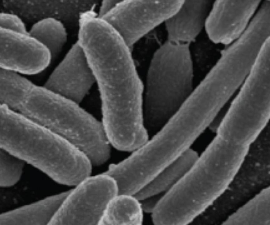
The term “bacillus” refers to a specific shape of bacteria that is cylindrical or rod-like. In clinical microbiology, identifying this shape is a primary step in the taxonomic classification of an unknown sample. While many bacilli are harmless or even beneficial to human health—such as the Lactobacillus species found in the gut—others are responsible for severe infections. The image shows a high-resolution scanning electron micrograph of several bacilli, highlighting their rounded ends and uniform structure.
The structural morphology of a bacillus is determined by its rigid cell wall and an internal cytoskeleton. The primary component of this wall is peptidoglycan, a complex polymer that provides the mechanical strength necessary to withstand high internal osmotic pressures. During growth, specialized proteins like MreB guide the synthesis of the cell wall, ensuring the bacterium maintains its rod shape as it elongates and prepares for division.
This specific shape offers several evolutionary advantages, particularly regarding movement and nutrient uptake. The elongated form of a bacillus increases its surface-area-to-volume ratio compared to spherical bacteria, which facilitates more efficient diffusion of gases and nutrients across the plasma membrane. This physiological efficiency is a key factor in the rapid growth rates observed in many bacterial populations.
Key characteristics of bacilli include:
- Diverse Arrangements: They can exist as single cells, pairs (diplobacilli), or long chains (streptobacilli).
- Motility: Many rod-shaped bacteria utilize flagella to move toward nutrient sources or away from toxic environments.
- Endospore Formation: Some genera, such as Bacillus and Clostridium, can form highly resistant dormant structures called endospores to survive extreme conditions.
- Variable Sizes: While typically 0.5 to 1.0 µm wide, their length can vary significantly depending on the species and environmental conditions.
Structural Anatomy and the Role of the Cell Wall
The anatomy of a bacillus is a marvel of biological engineering. Beneath the cell wall lies the plasma membrane, which regulates the transport of molecules and serves as the site for crucial metabolic processes like cellular respiration. Because prokaryotes lack mitochondria, the enzymes for ATP production are embedded directly into the plasma membrane. This streamlined design allows bacilli to respond rapidly to changes in their chemical surroundings.
The physical dimensions of these rods are not accidental. Research suggests that the rod shape is particularly effective for swimming in liquid environments. The longitudinal axis of the cell helps reduce drag, allowing motile bacilli to navigate through mucosal layers or aquatic habitats with greater speed and precision than their spherical counterparts. This motility is often a critical factor in the pathogenesis of bacterial infections, as it allows organisms to reach and colonize specific target tissues within a host.
Growth, Division, and Clinical Diagnostics
Bacilli reproduce through a process called binary fission, where the cell elongates to nearly twice its original size before splitting into two identical daughter cells. This process is highly regulated; the cell must precisely coordinate DNA replication with the synthesis of new membrane and wall material. In a laboratory setting, the way these cells stay attached after division provides diagnostic clues. For example, the formation of long chains is a hallmark of certain species that can be identified under a microscope following a Gram stain.
In a medical context, the distinction between different types of bacilli is vital for selecting the appropriate treatment. Gram-positive bacilli possess a thick layer of peptidoglycan, while Gram-negative bacilli have a thinner layer and an additional outer membrane. This structural difference determines the bacterium’s susceptibility to various antibiotics. For instance, some medications are specifically designed to penetrate the outer membrane of Gram-negative rods, while others target the cell wall synthesis of Gram-positive organisms.
The intricate design and physiological adaptability of bacilli illustrate the successful evolutionary strategies of prokaryotic life. From their efficient nutrient absorption to their specialized survival mechanisms like spore formation, these rod-shaped organisms continue to be a focal point of medical research. By advancing our understanding of bacterial anatomy, we enhance our ability to combat infectious diseases and harness the beneficial properties of these microscopic powerhouses.

