The axis bone, or second cervical vertebra, is a pivotal structure in the neck, enabling a wide range of head movements. This article provides a detailed look at its superior and anterior views, shedding light on the key anatomical features that define its role in spinal stability and motion.
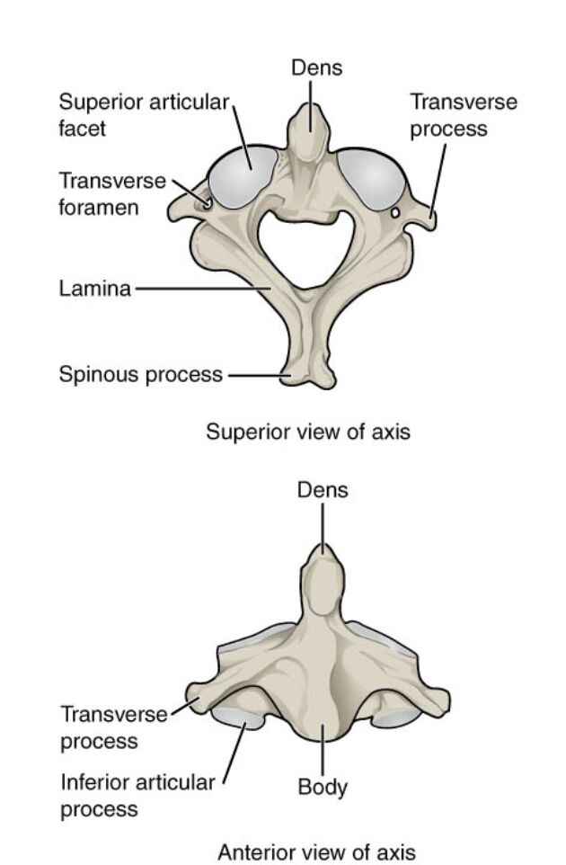
Dens: The dens, also known as the odontoid process, is a unique upward projection from the axis body that acts as a pivot for head rotation. It articulates with the atlas, allowing the head to turn side to side, and is critical for the atlantoaxial joint’s function.
Superior articular facet: The superior articular facet is a smooth surface on the axis that connects with the atlas above, facilitating smooth rotational movement. It plays a vital role in stabilizing the upper cervical spine during head motion.
Transverse foramen: The transverse foramen is a hole in the transverse process through which the vertebral artery and vein pass to supply blood to the brain. This structure is essential for maintaining adequate circulation to the head and neck.
Transverse process: The transverse process extends laterally from the axis, serving as an attachment point for muscles and ligaments that support neck movement. It also houses the transverse foramen, contributing to vascular support.
Lamina: The lamina forms the posterior portion of the vertebral arch, connecting the transverse and spinous processes. It protects the spinal cord and provides structural integrity to the axis.
Spinous process: The spinous process is a bony projection at the back of the axis, offering an attachment site for muscles and ligaments that aid in head and neck extension. It also serves as a landmark for surgical and diagnostic procedures.
Inferior articular process: The inferior articular process extends downward to articulate with the third cervical vertebra, ensuring continuity of the spinal column. It helps distribute loads and maintain alignment in the cervical spine.
Body: The body is the thick, weight-bearing anterior portion of the axis, providing a foundation for the dens and supporting the vertebral structure. It absorbs compressive forces and contributes to overall spinal stability.
Anatomical Overview of the Axis Bone
The axis bone stands out due to its distinctive features visible in the superior and anterior views. The dens is a defining characteristic, enabling the head’s rotational capacity.
- The superior articular facet allows for smooth interaction with the atlas, ensuring fluid movement.
- The transverse foramen supports critical vascular pathways, highlighting its role beyond skeletal support.
- The lamina and spinous process form a protective and stabilizing posterior framework.
- The body anchors the axis, distributing weight effectively across the cervical spine.
This combination of elements makes the axis a cornerstone of neck anatomy.
Functional Roles and Stability
The axis bone’s design supports both mobility and stability in the upper neck. The dens serves as the pivot point for the atlantoaxial joint, a key factor in head rotation.
- The superior articular facet and inferior articular process ensure proper alignment with adjacent vertebrae.
- The transverse process provides leverage for muscle action, enhancing neck flexibility.
- The lamina shields the spinal cord, while the spinous process aids in muscle attachment.
- The body bears the brunt of compressive forces, maintaining structural integrity.
These features work in harmony to support a wide range of motions.
Clinical Relevance and Movement
Understanding the axis bone’s anatomy has significant clinical implications. The dens’s position and stability are crucial for preventing dislocations or fractures in the upper cervical spine.
- The transverse foramen’s proximity to the vertebral artery makes it a focus in cases of vascular compromise.
- The superior articular facet’s articulation with the atlas is vital for diagnosing rotational injuries.
- The spinous process serves as a palpable landmark for assessing neck alignment.
- The inferior articular process and body contribute to load-bearing capacity, influencing spinal health.
This knowledge aids in diagnosing and treating cervical spine conditions effectively.
The axis bone, with its unique dens and robust structure, is essential for head movement and spinal support. The superior articular facet, transverse foramen, and lamina enhance its role in rotation and protection, while the spinous process, inferior articular process, and body provide stability and load distribution. Exploring these features deepens appreciation for the axis’s function and underscores the importance of preserving its integrity for overall neck health.

