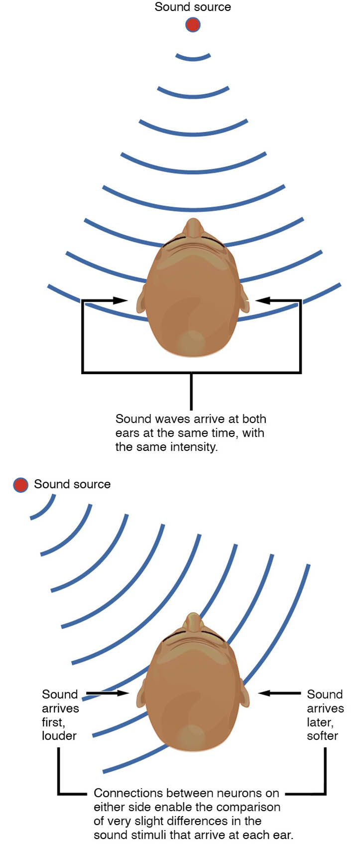
The auditory system’s ability to locate sound in the horizontal plane relies on intricate processing within the brain stem, enabling us to pinpoint the source of sounds in our environment. This image illustrates the medullary nuclei and their neural connections, showcasing how interaural time and intensity differences are analyzed to achieve precise sound localization.
Cochlear nucleus The cochlear nucleus is the first relay station in the auditory pathway, receiving input from the cochlea via the auditory nerve. It processes initial sound signals and sends them to higher auditory centers for further analysis.
Superior olivary complex The superior olivary complex integrates inputs from both ears, comparing interaural time differences to localize sound. It plays a key role in binaural processing, enhancing the brain’s ability to determine sound direction.
Medial superior olive (MSO) The medial superior olive (MSO) specializes in detecting interaural time differences, crucial for localizing low-frequency sounds. It contains neurons that compare the timing of sound arrival between the two ears.
Lateral superior olive (LSO) The lateral superior olive (LSO) focuses on interaural intensity differences, aiding in the localization of high-frequency sounds. It adjusts its response based on the relative loudness at each ear.
Trapezoid body The trapezoid body is a fiber bundle that connects the cochlear nuclei of both sides, facilitating communication between the auditory pathways. It supports the relay of signals to the superior olivary complex for binaural processing.
Inferior colliculus The inferior colliculus serves as a major integration center in the midbrain, receiving input from the superior olivary complex. It refines sound localization data and relays it to the thalamus and auditory cortex.
Medial geniculate nucleus (MGN) The medial geniculate nucleus (MGN) in the thalamus acts as a relay station, processing auditory information from the inferior colliculus. It forwards these signals to the auditory cortex for conscious perception.
Auditory cortex The auditory cortex, located in the temporal lobe, interprets the processed sound localization data from the MGN. It enables the conscious awareness and understanding of sound direction and characteristics.
Anatomy of Auditory Brain Stem Pathways
The brain stem houses critical nuclei that process sound localization, forming a network that connects the ears to the brain. This image highlights the anatomical layout of these structures in the auditory system.
- The cochlear nucleus receives auditory nerve fibers, marking the start of central processing.
- The superior olivary complex integrates bilateral inputs, enhancing spatial hearing.
- The medial superior olive (MSO) and lateral superior olive (LSO) specialize in timing and intensity cues.
- The trapezoid body serves as a bridge, linking the two sides of the auditory pathway.
- The inferior colliculus consolidates information in the midbrain for further relay.
- The medial geniculate nucleus (MGN) and auditory cortex complete the pathway to conscious perception.
- These structures are interconnected by well-defined neural tracts, ensuring efficient signal transmission.
Physiology of Sound Localization
Sound localization depends on the brain stem’s ability to analyze interaural differences, processed through specialized nuclei. This diagram illustrates the physiological mechanisms behind spatial hearing.
- The cochlear nucleus encodes initial sound frequencies and sends them to the superior olivary complex.
- The medial superior olive (MSO) compares the time delay of sound arrival, key for low frequencies.
- The lateral superior olive (LSO) assesses intensity differences, critical for high-frequency localization.
- The trapezoid body facilitates cross-talk between ears, supporting binaural comparison.
- The inferior colliculus integrates these cues, refining the spatial map.
- The medial geniculate nucleus (MGN) and auditory cortex process the final localization data.
- This system allows rapid adjustments to dynamic sound sources in the environment.
Role of the Superior Olivary Complex
The superior olivary complex is central to binaural processing, comparing inputs from both ears. Its subdivisions enhance the precision of sound localization.
- The medial superior olive (MSO) detects microsecond differences in sound arrival times.
- The lateral superior olive (LSO) evaluates amplitude disparities, aiding high-frequency localization.
- These nuclei work together to create a three-dimensional sound map.
- The complex receives inhibitory and excitatory inputs, fine-tuning its response.
- Damage to this area can impair directional hearing, though this image shows normal anatomy.
- Its location in the medulla optimizes early auditory processing.
Role of Higher Auditory Centers
The inferior colliculus, medial geniculate nucleus (MGN), and auditory cortex refine and interpret localization data. These structures complete the auditory pathway to conscious perception.
- The inferior colliculus integrates inputs from both olives, adding depth to localization.
- The medial geniculate nucleus (MGN) relays processed signals to the auditory cortex.
- The auditory cortex maps sound direction, combining cues with other sensory input.
- These centers adapt to changes in sound source position over time.
- Cross-modal interactions with vision enhance localization accuracy.
- Lesions here can lead to sound localization deficits, but this diagram depicts healthy structure.
Clinical Relevance of Auditory Localization
Understanding the brain stem mechanisms of sound localization supports the diagnosis and management of auditory disorders. This image provides a baseline for exploring related conditions.
- Damage to the cochlear nucleus can result from acoustic neuroma, affecting initial sound processing.
- Superior olivary complex dysfunction may cause difficulties in localizing sound sources.
- The medial superior olive (MSO) impairment can lead to poor low-frequency localization.
- The lateral superior olive (LSO) issues may reduce high-frequency directional accuracy.
- Inferior colliculus lesions can disrupt integrated auditory processing.
- Hearing aids and cochlear implants aim to compensate for pathway deficits.
- Auditory brainstem response (ABR) testing assesses these structures’ function.
In conclusion, the auditory brain stem mechanisms depicted in this image reveal a sophisticated system for sound localization. By analyzing interaural time and intensity differences through the cochlear nucleus, superior olivary complex, and higher centers, the brain accurately places sounds in space, offering a fascinating glimpse into auditory neuroscience.

