Atherosclerosis is a chronic condition marked by the accumulation of fatty, calcified plaques within artery walls, which can lead to serious cardiovascular complications. This diagram illustrates the process and impact of plaque formation, offering a visual representation of how it narrows and damages arteries over time. Exploring this image provides essential insights into the anatomy and progression of atherosclerosis, aiding in the recognition of its health implications.
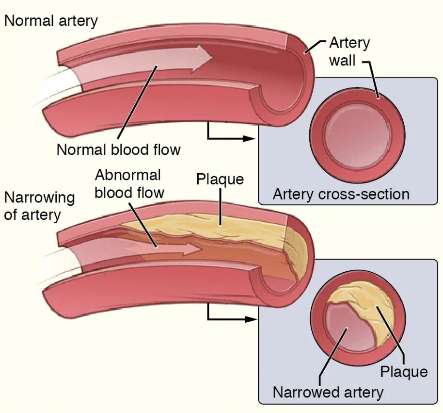
Artery wall: The artery wall is a multi-layered structure comprising the intima, media, and adventitia, designed to maintain elasticity and support blood flow. In atherosclerosis, this wall thickens due to plaque buildup, reducing its flexibility and increasing the risk of rupture.
Plaque: Plaque is a buildup of fatty substances, cholesterol, calcium, and cellular debris that forms within the artery wall, creating a hard, irregular deposit. This accumulation narrows the artery’s lumen, obstructing blood flow and potentially leading to clot formation or blockages.
Lumen: The lumen is the inner passageway of the artery where blood flows, normally wide and unobstructed to ensure adequate circulation. In atherosclerosis, the lumen becomes constricted by plaque, limiting oxygen delivery to tissues and organs.
Anatomical Structure of Atherosclerosis
The artery’s internal anatomy undergoes significant changes in atherosclerosis, and this diagram provides a detailed view of these transformations. Understanding the affected areas helps in grasping the disease’s impact on vascular health.
- The artery wall serves as the foundation where plaque begins to develop.
- The plaque’s growth disrupts the smooth inner lining, initiating the disease process.
- The lumen’s narrowing reflects the cumulative effect of plaque accumulation.
- This progression often starts with endothelial damage, a precursor to further complications.
The diagram highlights the artery’s vulnerability to these changes.
Physiological Impact and Symptoms
Atherosclerosis alters blood flow and oxygen supply, leading to a range of physiological effects. The image illustrates how these changes manifest within the artery.
- The artery wall’s thickening increases resistance, elevating blood pressure in affected areas.
- The plaque’s presence can trigger inflammation, heightening the risk of thrombosis.
- The lumen’s reduction may cause chest pain or leg cramps, signaling reduced blood flow.
- Symptoms vary by location, with coronary arteries often linked to angina or heart attack.
Early detection through imaging can mitigate severe outcomes.
Causes and Risk Factors
The development of atherosclerosis involves multiple contributing factors that drive plaque formation. Identifying these aids in prevention and management strategies.
- High levels of low-density lipoprotein cholesterol promote plaque buildup in the artery wall.
- Hypertension damages the endothelium, accelerating atherosclerosis progression.
- Smoking and diabetes increase oxidative stress, worsening plaque development.
- Genetic factors, such as family history, heighten susceptibility to this condition.
A heart-healthy diet and regular exercise can help reduce these risks.
Diagnosis and Treatment Options
Diagnosing and managing atherosclerosis requires a comprehensive approach based on its stage and symptoms. Advanced techniques guide therapeutic decisions.
- Angiography visualizes the lumen to assess the extent of plaque obstruction.
- Statins lower cholesterol, slowing plaque growth within the artery wall.
- Angioplasty or stenting widens the lumen, restoring blood flow in narrowed arteries.
- Bypass surgery may be necessary for severe cases involving extensive plaque.
Regular check-ups with lipid profiles monitor disease progression.
Clinical Relevance and Long-Term Outlook
Understanding the implications of atherosclerosis is crucial for long-term vascular health. The condition’s effects depend on the degree of arterial involvement.
- The artery wall’s integrity affects its ability to adapt to changing blood flow demands.
- The plaque’s stability influences the risk of acute events like myocardial infarction.
- The lumen’s patency post-treatment determines exercise tolerance and organ function.
- Lifestyle changes, such as quitting smoking, improve outcomes and quality of life.
Ongoing management with medication supports vascular resilience.
Conclusion
This diagram of atherosclerosis provides a detailed view of the artery wall, plaque, and lumen, illustrating the disease’s impact on arterial health. By depicting the buildup of fatty, calcified deposits and their effect on blood flow, it underscores the importance of early detection and intervention to prevent complications. This understanding equips individuals with the knowledge to address atherosclerosis effectively, promoting better cardiovascular outcomes.

