The arteries supplying the head and neck play an essential role in delivering oxygenated blood to the brain, face, and upper structures, ensuring vital functions like cognition and sensory processing. This diagram illustrates the pathways of the common carotid, external carotid, internal carotid, vertebral, and subclavian arteries, highlighting their intricate network and anatomical significance.
Common carotid artery This major artery arises from the brachiocephalic trunk on the right and the aortic arch on the left, supplying the head and neck. It bifurcates into the external and internal carotid arteries to distribute blood effectively.
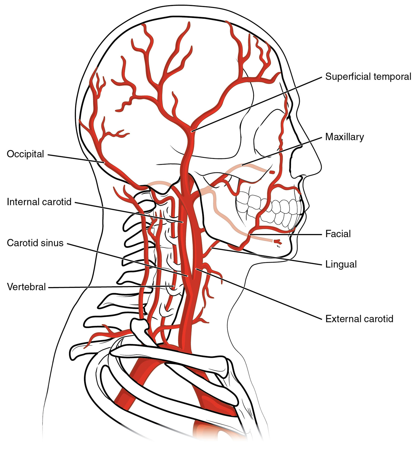
External carotid artery Remaining superficial, this artery supplies blood to the face, scalp, and parts of the neck. It gives rise to multiple branches, including the facial and superficial temporal arteries.
Internal carotid artery This artery extends deeper, initially forming the carotid sinus for pressure regulation. It passes through the carotid canal and foramen lacerum to supply the brain with oxygenated blood.
Carotid sinus Located at the bifurcation of the common carotid, this dilated area contains baroreceptors. It monitors blood pressure and helps regulate cardiovascular responses.
Carotid canal This bony passage in the temporal bone guides the internal carotid artery toward the skull. It protects the artery as it approaches the brain.
Carotid foramen This opening connects the carotid canal to the cranial cavity. It allows the internal carotid artery to enter the skull safely.
Foramen lacerum This irregular opening at the skull base permits the internal carotid artery to emerge into the cranium. It serves as a critical entry point for brain circulation.
Vertebral artery Branching from the subclavian artery, it travels through the transverse foramina of cervical vertebrae. It enters the skull via the vertebral foramen to supply the brain and spinal cord.
Subclavian artery This artery originates from the brachiocephalic trunk or aortic arch, supplying the upper limb and parts of the head. It continues as the axillary artery toward the arm.
Axillary artery Extending from the subclavian artery, it provides blood to the shoulder and upper arm. It supports the muscles and tissues of the upper limb.
Transverse foramen These openings in the cervical vertebrae protect the vertebral artery’s path. They guide the artery upward toward the skull base.
Vertebral foramen Located at the skull base, this foramen allows the vertebral artery to enter the cranial cavity. It facilitates blood supply to the posterior brain regions.
Anatomy of the Common Carotid and Its Branches
The common carotid artery serves as the primary conduit for head and neck circulation. Its bifurcation into external and internal branches ensures comprehensive coverage.
- The right common carotid arises from the brachiocephalic artery, while the left stems from the aortic arch.
- The external carotid artery supplies superficial structures like the face and scalp.
- The internal carotid artery dives deeper to nourish the brain.
- The carotid sinus at the bifurcation acts as a pressure sensor.
- This network adapts to varying blood demands during activity or rest.
Pathway of the Internal Carotid Artery
The internal carotid artery follows a complex route to reach the brain. Its passage through cranial structures is meticulously designed for protection.
- The carotid sinus initiates pressure regulation before the artery enters the carotid canal.
- The carotid canal shields the artery as it ascends through the temporal bone.
- The carotid foramen and foramen lacerum guide it into the cranial cavity.
- Once inside, it supplies the anterior and middle cerebral arteries.
- Blockages here can lead to severe neurological deficits.
Role of the Vertebral Artery
The vertebral artery provides an alternative route for brain blood supply. Its journey through the cervical spine is both protective and functional.
- Originating from the subclavian artery, it passes through the transverse foramen of cervical vertebrae.
- The vertebral foramen at the skull base allows entry for posterior circulation.
- It joins the opposite vertebral artery to form the basilar artery.
- This artery nourishes the brainstem and cerebellum.
- Its path makes it vulnerable to compression or trauma.
Connection to the Upper Limb
The subclavian artery and its continuation as the axillary artery link head and neck circulation to the arm. This transition supports upper body function.
- The subclavian artery branches to supply the vertebral artery and upper limb.
- The axillary artery extends into the arm, feeding shoulder muscles.
- This connection ensures coordinated blood flow during movement.
- Narrowing of the subclavian can reduce arm or brain perfusion.
- The system adjusts flow based on physical demands.
Clinical Relevance of Head and Neck Arteries
Understanding these arteries aids in diagnosing and treating vascular issues. Their anatomy is critical for medical interventions.
- Stenosis in the internal carotid artery can cause strokes.
- Carotid sinus hypersensitivity may lead to fainting episodes.
- Vertebral artery dissection is a risk in neck trauma.
- Subclavian steal syndrome affects blood flow to the brain and arm.
- Imaging like Doppler ultrasound assesses these arteries for disease.
The arteries supplying the head and neck, from the common carotid artery to the vertebral artery, form a vital network delivering oxygenated blood. Their intricate pathways ensure the brain and upper structures thrive, providing a foundation for exploring circulatory health and addressing related conditions.

