The arteries of the thoracic and abdominal regions play a crucial role in delivering oxygenated blood to the chest, abdomen, and their associated organs, ensuring vital physiological processes. This diagram illustrates the thoracic aorta and its visceral and parietal branches, providing a detailed map of how blood is distributed to support digestion, respiration, and other functions.
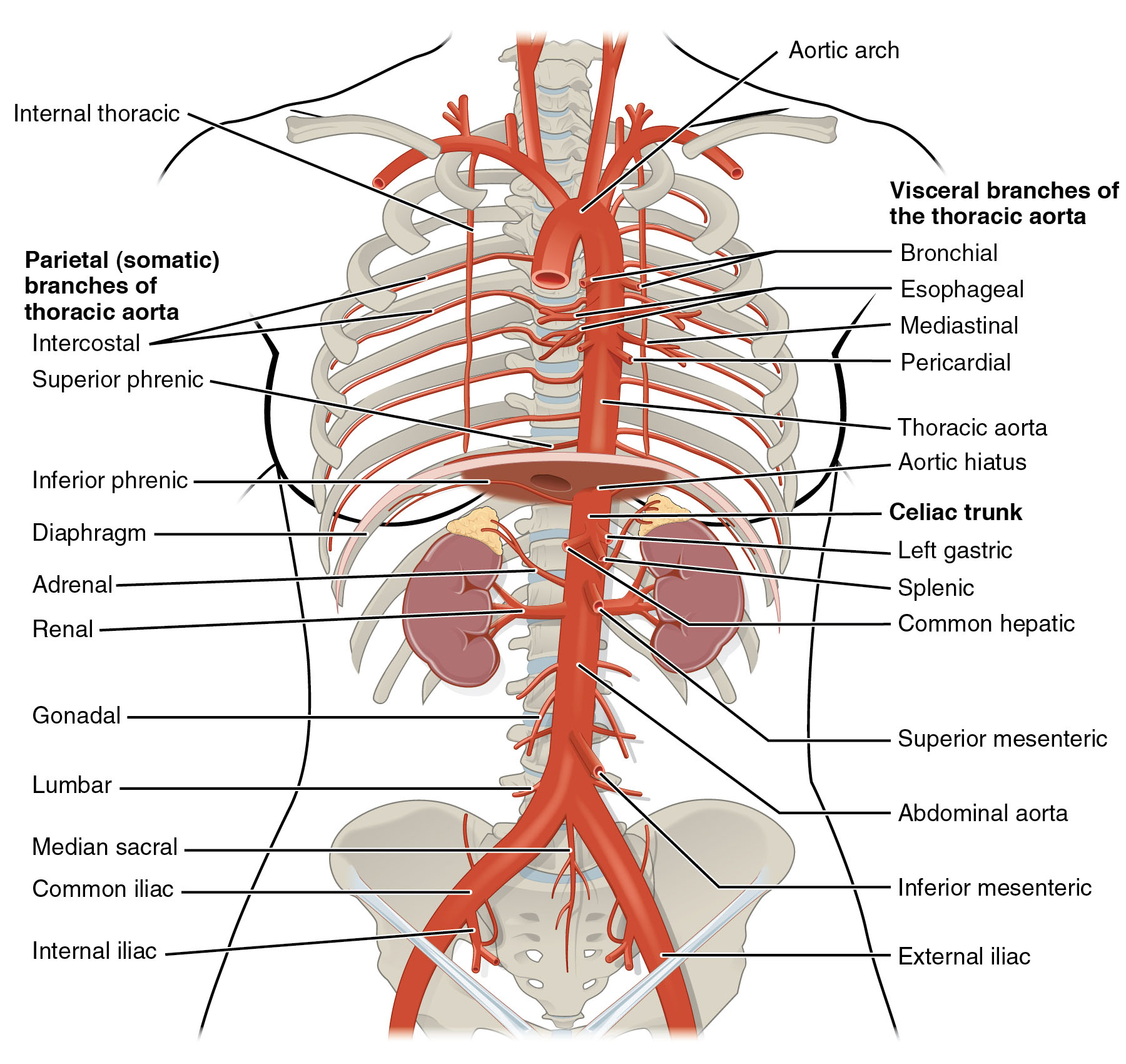
Thoracic aorta This segment of the descending aorta runs through the chest, supplying blood to thoracic organs. It gives rise to both visceral and parietal branches to meet regional demands.
Visceral branches These arteries supply the internal organs of the thoracic cavity, such as the lungs and esophagus. They ensure oxygen and nutrients reach tissues critical for respiration and digestion.
Pericardial arteries Branching from the thoracic aorta, they supply blood to the pericardium surrounding the heart. They support the heart’s protective sac, aiding in its structural integrity.
Bronchial arteries These arteries arise from the thoracic aorta to supply the lungs with blood. They nourish the bronchial tree and support pulmonary tissue function.
Esophageal arteries Originating from the thoracic aorta, they provide blood to the esophagus. They ensure the esophagus remains healthy for food passage to the stomach.
Parietal branches These arteries supply the chest wall and diaphragm, supporting structural and muscular components. They deliver blood to the intercostal muscles and other thoracic structures.
Posterior intercostal arteries Branching from the thoracic aorta, they supply the intercostal muscles and chest wall. They play a key role in respiration by supporting rib movement.
Subcostal arteries These arteries, also from the thoracic aorta, supply the lower chest wall and upper abdomen. They provide blood to the diaphragm and adjacent tissues.
Abdominal aorta Continuing from the thoracic aorta, this segment runs through the abdomen, supplying major organs. It gives rise to arteries like the celiac trunk and renal arteries.
Celiac trunk This branch of the abdominal aorta supplies the stomach, liver, and spleen. It ensures blood flow for digestion, detoxification, and nutrient storage.
Superior mesenteric artery Arising from the abdominal aorta, it feeds the small intestine and part of the large intestine. It supports nutrient absorption and gastrointestinal health.
Renal arteries These arteries branch from the abdominal aorta to supply the kidneys. They deliver blood for filtration, waste removal, and blood pressure regulation.
Inferior mesenteric artery Originating from the abdominal aorta, it supplies the lower large intestine. It ensures blood flow for waste elimination and lower digestive function.
Common iliac arteries Splitting from the abdominal aorta, they supply the pelvis and lower limbs. They support leg movement and pelvic organ perfusion with oxygenated blood.
Anatomy of the Thoracic Aorta
The thoracic aorta serves as a vital conduit for blood distribution in the chest region. Its branching pattern supports a variety of thoracic structures.
- The visceral branches, like the bronchial and esophageal arteries, nourish internal organs.
- Parietal branches, such as the posterior intercostal arteries, feed the chest wall.
- The aorta’s elastic walls absorb pressure from the heart’s contractions.
- Its location allows it to adapt to respiratory movements.
- This segment’s health is crucial for lung and heart function.
Role of Visceral and Parietal Branches
Visceral branches and parietal branches enhance the thoracic aorta’s reach. Each type targets specific anatomical needs with precision.
- Pericardial arteries support the heart’s protective layer, preventing friction.
- Bronchial arteries ensure lung tissue remains oxygenated and functional.
- Esophageal arteries maintain esophageal integrity for swallowing.
- Posterior intercostal arteries aid in chest expansion during breathing.
- These branches collectively sustain thoracic vitality.
Anatomy of the Abdominal Aorta
The abdominal aorta continues the aorta’s role, delivering blood to the lower body. Its branches are tailored to abdominal organ demands.
- The celiac trunk feeds the upper digestive organs for metabolism.
- Superior mesenteric artery supports the intestines’ absorptive capacity.
- Renal arteries ensure kidney filtration and hormone production.
- Inferior mesenteric artery sustains the lower digestive tract.
- The common iliac arteries extend circulation to the pelvis and legs.
Physiological Importance of These Arteries
The arteries from the thoracic and abdominal aorta maintain blood pressure and organ perfusion. Their function is integral to systemic health.
- The thoracic aorta’s branches adjust flow based on respiratory needs.
- Abdominal branches respond to digestive and excretory demands.
- Elasticity in these arteries smooths out pressure variations.
- Oxygen delivery supports cellular metabolism across regions.
- This network prevents ischemia in vital organs during stress.
Clinical Relevance of Thoracic and Abdominal Arteries
Knowledge of these arteries aids in diagnosing and treating vascular conditions. Their anatomy guides medical and surgical strategies.
- Aneurysms in the thoracic aorta can compress nearby structures.
- Blockages in renal arteries may lead to hypertension or kidney failure.
- Superior mesenteric artery occlusion can cause intestinal ischemia.
- Abdominal aortic aneurysms are a significant health concern.
- Imaging like CT angiography maps these arteries for intervention.
The thoracic aorta and abdominal aorta, with their visceral and parietal branches, form a robust network delivering oxygenated blood to the chest and abdomen. Their intricate design ensures organs like the lungs, kidneys, and intestines thrive, providing a foundation for understanding circulatory health and addressing related challenges.

