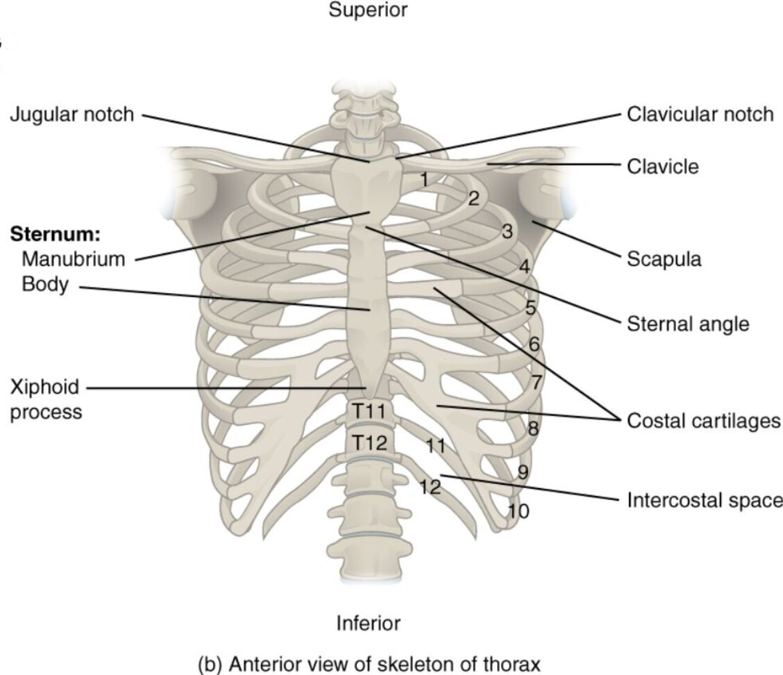
The anterior view of the thorax skeleton offers a comprehensive look at the chest’s bony framework, essential for protecting vital organs like the heart and lungs. This region’s structure supports respiration, provides attachment points for muscles, and maintains the body’s upright posture, making it a cornerstone of anatomical study.
Labels Introduction
Jugular notch: The jugular notch is a prominent indentation at the superior border of the manubrium, easily palpable and serving as a key landmark for clinical examinations. It lies between the clavicular notches and is used to locate the internal jugular vein and other structures.
Clavicular notch: The clavicular notch is a paired depression on either side of the manubrium where the clavicles articulate, forming part of the sternoclavicular joint. This notch ensures stability and mobility of the shoulder girdle.
Clavicle: The clavicle, or collarbone, is a slender, S-shaped bone extending laterally from the sternum to the scapula, providing support to the shoulder. It acts as a strut, transmitting forces from the upper limb to the axial skeleton.
Scapula: The scapula, or shoulder blade, is a flat, triangular bone located on the posterior thorax, partially visible anteriorly, and serves as an attachment site for muscles of the arm and chest. It facilitates a wide range of shoulder movements.
Sternal angle: The sternal angle, or angle of Louis, is a transverse ridge where the manubrium joins the body of the sternum, marking the level of the second rib. It is a critical landmark for identifying rib levels and auscultating heart sounds.
Costal cartilages: The costal cartilages are flexible, hyaline cartilage structures connecting the ribs to the sternum, allowing the rib cage to expand during breathing. They provide elasticity and support to the thoracic cage.
Intercostal space: The intercostal space is the area between adjacent ribs, housing intercostal muscles, nerves, and blood vessels that aid in respiration. These spaces are vital for maintaining the flexibility and movement of the rib cage.
Sternum: Manubrium: The manubrium is the uppermost part of the sternum, a broad, quadrangular structure that articulates with the clavicles and first two ribs. It forms the superior boundary of the thoracic cavity and protects underlying structures.
Sternum: Body: The body of the sternum is the longest middle portion, formed by the fusion of sternebrae, and articulates with the costal cartilages of ribs 2 through 7. It provides a central anchor for the rib cage and protects the heart.
Xiphoid process: The xiphoid process is the small, cartilaginous lower end of the sternum that ossifies with age, serving as an attachment point for the diaphragm and abdominal muscles. It can be a landmark for cardiopulmonary resuscitation.
Detailed Anatomical Overview
Structure of the Thorax Skeleton
The anterior thorax skeleton forms a protective cage around the thoracic cavity, safeguarding the heart, lungs, and major blood vessels. The jugular notch and clavicular notch define the superior boundary, offering accessible points for medical procedures. This framework supports the upper body and facilitates breathing through its dynamic structure.
- The clavicle connects the sternum to the scapula, stabilizing the shoulder while allowing arm movement.
- The scapula anchors muscles like the trapezius and deltoid, contributing to upper limb mobility.
- The sternal angle aligns with the second costal cartilage, aiding in locating the second intercostal space for chest procedures.
Functional Roles and Clinical Significance
The thorax’s bony structure enables expansion and contraction during respiration, driven by the costal cartilages and intercostal spaces. The sternum: manubrium protects the great vessels, such as the aorta, while the sternum: body supports the rib cage’s lateral expansion. These elements are crucial for maintaining thoracic integrity.
- The jugular notch is used to assess central venous pressure by locating the jugular vein.
- The clavicular notch can be involved in sternoclavicular dislocations, requiring careful evaluation.
- The sternal angle serves as a reference for inserting chest tubes or performing pericardiocentesis.
Muscular and Neural Support
The intercostal spaces contain the external and internal intercostal muscles, which assist in elevating and depressing the ribs during breathing. The xiphoid process anchors the diaphragm, a primary muscle of respiration, and the rectus abdominis. This coordination ensures efficient oxygen intake and carbon dioxide expulsion.
- The costal cartilages allow the ribs to move, accommodating lung expansion.
- The scapula supports the rotator cuff muscles, essential for arm rotation and lifting.
- The sternum: body provides attachment for the pectoralis major, aiding in arm flexion.
Clinical Applications and Anatomical Insights
The thorax skeleton is a frequent focus in trauma and surgery, with the clavicle being prone to fractures due to its position. The sternal angle helps identify the level of the pulmonary artery, guiding catheter placement. Understanding these landmarks enhances diagnostic accuracy and procedural success.
- The jugular notch can indicate tracheal deviation in cases of mediastinal shift.
- The manubrium may be compressed during CPR, requiring precise force application.
- The xiphoid process can be a site of injury, leading to rare conditions like xiphodynia.
The anterior thorax skeleton is a marvel of engineering, balancing protection and flexibility. Its labeled components work together to support vital functions, making it a key area for anatomical study and clinical practice. Mastery of this region improves the ability to address thoracic conditions with precision.
Conclusion
The anterior view of the thorax skeleton reveals a robust structure that shields essential organs while enabling movement and respiration. This anatomical region’s intricate design supports daily activities and serves as a critical reference in medical interventions. Exploring its components deepens appreciation for its role in maintaining overall health and well-being.

