The ankle joint is a critical structure in the human body, facilitating movement and stability with its intricate network of bones and ligaments. This article delves into the anatomical details of the talocrural and subtalar joints, providing a comprehensive overview of their components and functions to enhance understanding of this vital area.
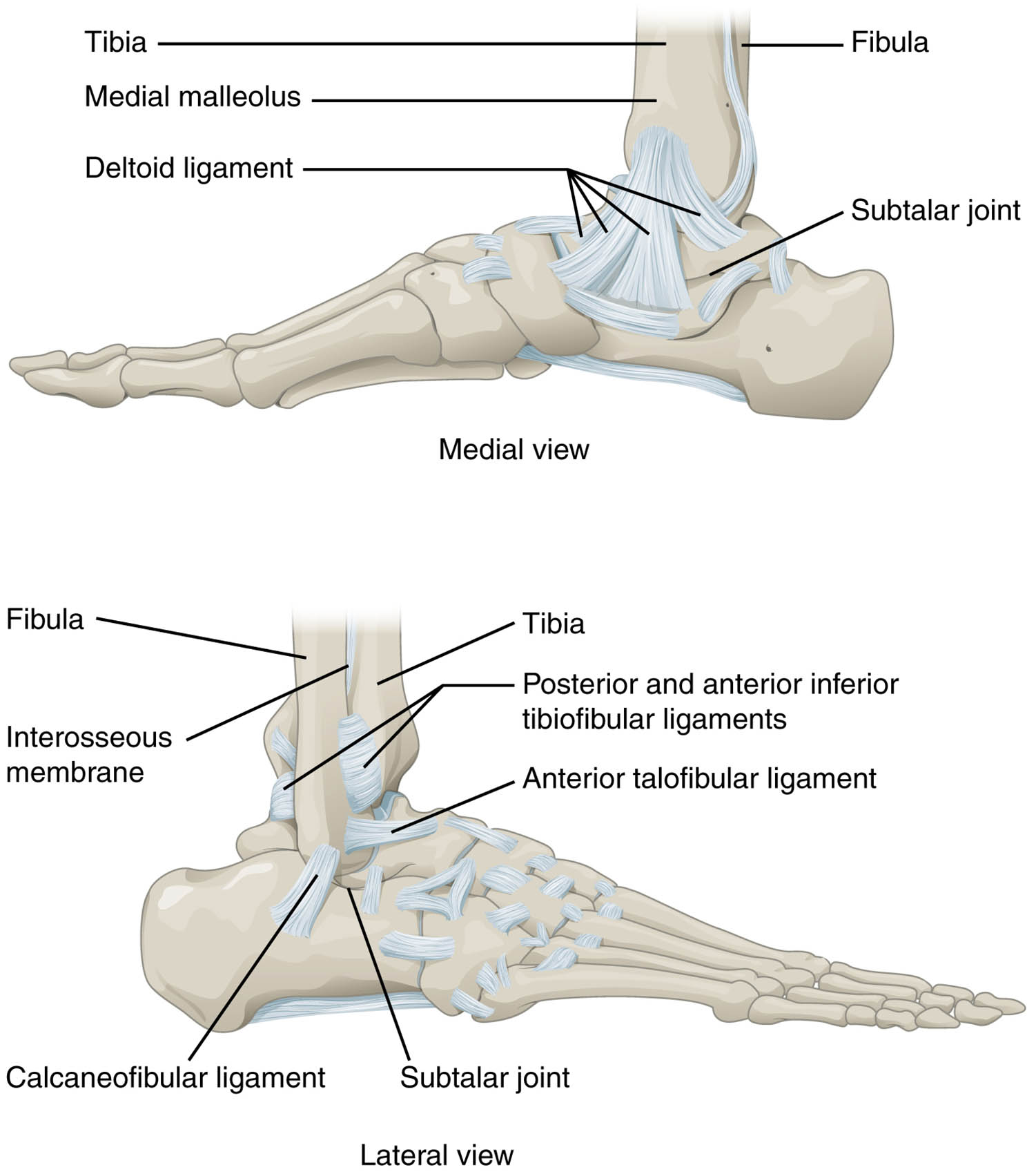
Tibia: The tibia, commonly known as the shinbone, is the larger and stronger of the two bones in the lower leg, providing primary support to the ankle joint. It articulates with the talus to form the medial portion of the talocrural joint, playing a key role in weight-bearing and stability.
Fibula: The fibula, a slender bone parallel to the tibia, contributes to the lateral stability of the ankle by forming the lateral malleolus. It works alongside the tibia to support the ankle and facilitate smooth movement during dorsiflexion and plantar flexion.
Medial malleolus: The medial malleolus is the bony prominence on the inner side of the ankle, formed by the distal end of the tibia. It serves as an attachment point for ligaments and helps stabilize the ankle against excessive inversion.
Deltoid ligament: The deltoid ligament is a strong, triangular ligament on the medial side of the ankle, connecting the tibia to the talus and calcaneus. It provides crucial support, resisting excessive eversion and protecting the ankle from lateral stress.
Subtalar joint: The subtalar joint, located between the talus and calcaneus, allows for inversion and eversion movements of the foot. This joint works in conjunction with other intertarsal joints to enhance foot mobility and adaptability.
Posterior and anterior inferior tibiofibular ligaments: These ligaments connect the tibia and fibula, stabilizing the distal tibiofibular syndesmosis. They play a vital role in maintaining the integrity of the ankle joint during weight-bearing activities.
Anterior talofibular ligament: The anterior talofibular ligament runs from the fibula to the talus, providing lateral stability to the ankle. It is commonly involved in ankle sprains due to its vulnerability during inversion injuries.
Calcaneofibular ligament: The calcaneofibular ligament extends from the fibula to the calcaneus, offering additional lateral support to the ankle. It helps prevent excessive inversion and contributes to the overall stability of the joint.
Interosseous membrane: The interosseous membrane is a fibrous sheet connecting the tibia and fibula along their length. It enhances the stability of the lower leg and serves as an attachment site for muscles and ligaments.
Detailed Anatomical Overview
Understanding the ankle joint begins with recognizing its role as a uniaxial hinge joint. The talocrural joint, formed by the tibia, fibula, and talus, primarily allows dorsiflexion and plantar flexion, essential for walking and running.
- The tibia and fibula form a mortise that encases the talus, ensuring a stable platform for movement.
- Ligaments such as the deltoid ligament and anterior talofibular ligament reinforce this structure, preventing excessive motion.
- The subtalar joint adds a layer of complexity, enabling side-to-side movements that adapt the foot to uneven surfaces.
This combination of joints and ligaments creates a dynamic system capable of withstanding significant stress.
Ligament Functions and Stability
Ligaments are the unsung heroes of ankle stability. Each plays a specific role in maintaining the joint’s integrity.
- The deltoid ligament is particularly robust, offering resistance against eversion forces that could otherwise destabilize the ankle.
- The anterior talofibular ligament and calcaneofibular ligament work together to limit inversion, a common mechanism in ankle injuries.
- The posterior and anterior inferior tibiofibular ligaments ensure the tibia and fibula remain aligned, critical for load distribution.
Together, these structures protect the ankle from sprains and other injuries, making them essential for daily mobility.
Clinical Relevance and Movement
The ankle’s design supports a range of motions critical for physical activity. The talocrural joint’s hinge action is complemented by the subtalar joint‘s ability to invert and evert the foot.
- Dorsiflexion and plantar flexion are powered by muscles like the tibialis anterior and gastrocnemius, coordinated with joint stability.
- The interosseous membrane aids in distributing forces between the tibia and fibula, reducing strain during movement.
- Proper alignment of the medial malleolus and lateral malleolus ensures efficient load transfer to the foot.
This biomechanical synergy underscores the ankle’s importance in locomotion and balance.
The ankle joint’s intricate anatomy, featuring the talocrural and subtalar joints, is a testament to its role in human mobility. Bones like the tibia and fibula, supported by ligaments such as the deltoid ligament and anterior talofibular ligament, create a resilient structure. The subtalar joint enhances flexibility, while the interosseous membrane and calcaneofibular ligament provide additional support. Understanding these components fosters appreciation for the ankle’s function and the need to protect it from injury, ensuring long-term health and activity.

