The male external genitalia plays a crucial role in both urinary and reproductive functions. Among its components, the penis is central, and its appearance can vary significantly depending on whether circumcision has been performed. This diagram specifically illustrates the external anatomy of a circumcised penis, highlighting its key features without the presence of the foreskin. Understanding these anatomical landmarks is fundamental for comprehending variations in male anatomy and related health discussions.
Detailed Anatomy of the Circumcised Penis
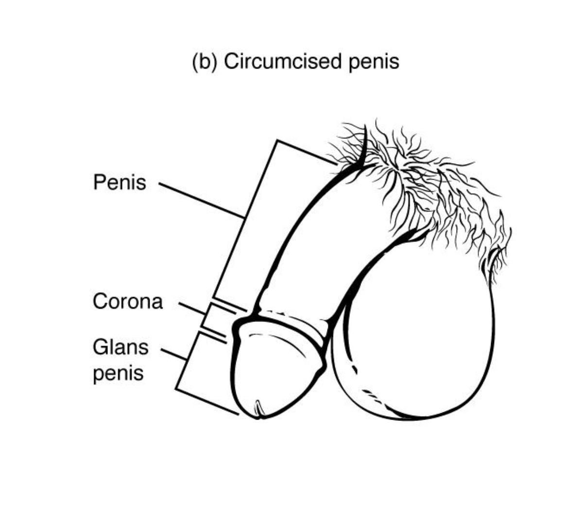
b) Circumcised penis: This image provides a visual representation of a penis that has undergone circumcision, a surgical procedure where the foreskin (prepuce) is removed. The defining characteristic is the exposed glans penis.
Penis: The primary external male copulatory organ, through which both urine and semen are expelled from the body. It consists of a shaft and the glans penis, and its internal structure involves erectile tissues.
Corona: The crown-like ridge located at the base of the glans penis, separating the glans from the shaft of the penis. It is an area known for its heightened sensitivity due to a rich supply of nerve endings.
Glans penis: The enlarged, rounded, and highly sensitive tip of the penis. In a circumcised individual, this structure is permanently exposed, distinguishing it from an uncircumcised penis where it is covered by the foreskin.
Understanding Circumcision and Penile Anatomy
The diagram presents a clear visual of the circumcised penis, a common anatomical variation resulting from a surgical procedure. Circumcision involves the removal of the prepuce, or foreskin, which is the retractable fold of skin that naturally covers the glans penis. This surgical modification permanently exposes the glans, revealing the distinct anatomical landmarks shown in the illustration.
The primary external components depicted are:
- The Penis: The main organ, responsible for urinary excretion and sexual function.
- The Corona: A prominent ridge that demarcates the glans from the shaft.
- The Glans Penis: The sensitive tip of the penis, which, in a circumcised state, is uncovered.
The practice of circumcision is widespread globally, driven by various cultural, religious, and medical considerations. From a medical perspective, it is sometimes performed to address conditions such as phimosis (a foreskin that is too tight to retract) or recurrent infections. Understanding the external anatomy post-circumcision is essential for healthcare professionals and individuals alike, as it influences hygiene practices, sexual sensation, and potential health considerations related to the exposed glans.
The Circumcised Penis: Anatomy and Clinical Relevance
The male external genitalia, particularly the penis, exhibits anatomical variations that are important in both clinical practice and public health discussions. One such variation is the circumcised penis, which results from a surgical procedure to remove the foreskin (prepuce). This diagram specifically illustrates the external features of a circumcised penis, highlighting the glans and corona. This anatomical presentation is prevalent in many parts of the world, influenced by a combination of cultural, religious, and medical factors.
The most defining characteristic of the circumcised penis is the permanent exposure of the glans penis, the highly sensitive, bulbous tip of the organ. In an uncircumcised individual, the glans is covered by the prepuce, a movable fold of skin. The absence of this foreskin in circumcised males means that the glans is continually exposed to air and friction, which can lead to a slight keratinization or toughening of its surface over time. Immediately posterior to the glans is the corona, a raised ridge that marks the boundary between the glans and the shaft of the penis. This area is particularly rich in nerve endings and plays a role in sexual sensation.
From a clinical standpoint, circumcision is performed for various reasons. In some cases, it is medically indicated to treat or prevent conditions such as phimosis (a foreskin that cannot be retracted), paraphimosis (a retracted foreskin that cannot be returned to its normal position), balanitis (inflammation of the glans), or recurrent urinary tract infections in infants. Religious and cultural traditions also significantly contribute to the prevalence of circumcision in many communities worldwide. Discussions surrounding circumcision often involve considerations of hygiene, potential effects on sexual sensation, and risks associated with the surgical procedure. While hygiene practices may differ, the fundamental anatomy of the circumcised penis, as depicted, remains consistent.
Understanding the specific anatomical landmarks of the circumcised penis, such as the glans and corona, is crucial for healthcare providers during examinations, for educating patients on hygiene, and for discussing potential health outcomes. The absence of the foreskin inherently alters some aspects of penile care and can influence sensitivity, though the precise degree of impact on sensation is a subject of ongoing research and individual variation. This visual representation serves as a foundational reference for appreciating this common anatomical presentation in male individuals.
Conclusion
This diagram of the circumcised penis offers a clear and concise depiction of its external anatomy, focusing on the exposed glans and the prominent corona. The absence of the foreskin is the defining characteristic, distinguishing it from an uncircumcised state. This anatomical understanding is important for various reasons, including clinical assessment, discussions on hygiene, and comprehending the diverse presentations of male external genitalia.

