The brain stem is a vital component of the central nervous system, serving as a conduit between the brain and spinal cord while regulating essential life-sustaining functions. This midsagittal view illustrates the brain stem’s three primary regions—the midbrain, pons, and medulla—highlighting their anatomical continuity and roles in motor control, sensory processing, and autonomic regulation. Understanding these structures provides key insights into neurological health and basic physiological processes.
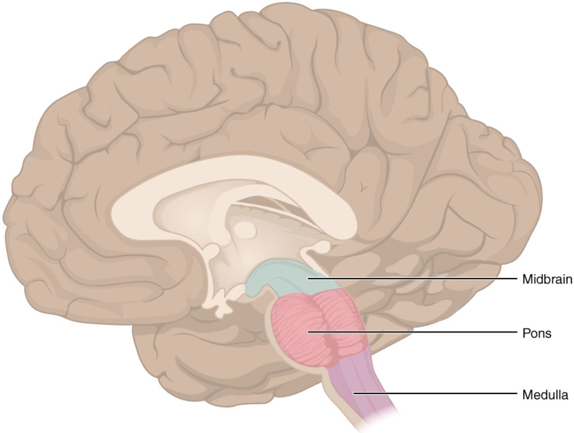
Midbrain
The midbrain, located superiorly in the brain stem, connects the forebrain to the hindbrain and houses critical nuclei for visual and auditory reflexes. It contains the substantia nigra, which produces dopamine essential for movement coordination, and the cerebral aqueduct, facilitating cerebrospinal fluid flow.
Pons
The pons, positioned between the midbrain and medulla, acts as a bridge for neural pathways connecting the cerebrum to the cerebellum, supporting coordinated movement and balance. It includes nuclei for cranial nerves V through VIII, influencing facial sensation, hearing, and equilibrium.
Medulla
The medulla, the most inferior part of the brain stem, regulates vital autonomic functions such as heart rate, respiration, and blood pressure through its reticular formation and cranial nerve nuclei. It transitions into the spinal cord, serving as a relay for sensory and motor tracts crossing at the pyramidal decussation.
Overview of Brain Stem Anatomy
The brain stem forms the core of the hindbrain, extending from the diencephalon to the spinal cord. Its compact structure integrates multiple neural pathways essential for survival.
- The brain stem measures approximately 7-8 cm in length, comprising about 2-3% of total brain mass yet critical for basic functions.
- It is divided into midbrain (mesencephalon), pons (metencephalon component), and medulla oblongata (myelencephalon).
- Vascular supply primarily from the vertebral and basilar arteries ensures constant perfusion, with the blood-brain barrier protecting against toxins.
- Embryologically, it develops from the rhombencephalon and mesencephalon, with neuronal migration establishing its layered organization.
Detailed Structure of the Midbrain
The midbrain serves as a transitional zone with distinct dorsal and ventral divisions. Its location allows integration of higher and lower neural signals.
- The tectum includes superior and inferior colliculi, processing visual (superior) and auditory (inferior) reflexes via tectospinal and tectobulbar tracts.
- The tegmentum contains the red nucleus for motor coordination and the periaqueductal gray for pain modulation and autonomic responses.
- Cranial nerves III and IV originate here, controlling eye movements and pupil constriction.
- Dopaminergic neurons in the substantia nigra project to the striatum, forming the nigrostriatal pathway crucial for voluntary movement.
Anatomy and Role of the Pons
The pons features prominent transverse fibers connecting to the cerebellum. Its bulbous shape reflects its role in relaying information.
- Pontine nuclei facilitate corticopontocerebellar tracts, enabling fine motor adjustments and learning.
- The reticular formation within the pons contributes to arousal, sleep-wake cycles, and respiratory rhythm generation.
- Cranial nerve V (trigeminal) handles mastication and facial sensation, while VI, VII, and VIII manage eye abduction, facial expression, and vestibulocochlear functions.
- The medial longitudinal fasciculus coordinates conjugate eye movements, linking to vestibular inputs.
Functions of the Medulla Oblongata
The medulla controls life-essential reflexes and autonomic outputs. Its elongated form transitions seamlessly into the spinal cord.
- Vital centers include the cardiac center for heart rate modulation via sympathetic and parasympathetic fibers, and the vasomotor center for vascular tone.
- The respiratory center, comprising dorsal and ventral groups, regulates breathing rhythm and depth in response to CO2 levels.
- Cranial nerves IX through XII originate here, influencing swallowing, speech, and tongue movement.
- Sensory tracts like the dorsal column-medial lemniscus decussate, while motor pyramidal tracts cross at the pyramids for contralateral control.
Integrated Physiology of the Brain Stem
These regions function cohesively to maintain homeostasis and coordinate responses. Their interconnected pathways ensure efficient signal transmission.
- Ascending reticular activating system (ARAS) from the brain stem promotes cortical arousal and consciousness.
- Descending tracts like rubrospinal and vestibulospinal modulate posture and balance.
- Neurotransmitters such as norepinephrine from locus coeruleus in the pons influence attention and stress responses.
- The brain stem’s role in reflex arcs, like the baroreceptor reflex, stabilizes blood pressure through glossopharyngeal and vagus nerves.
Clinical Relevance and Neural Pathways
Knowledge of brain stem anatomy informs diagnostic and therapeutic approaches. Its vulnerability underscores the need for precise imaging.
- MRI and CT scans visualize brain stem structures, aiding in identifying infarcts or hemorrhages.
- The midbrain’s Weber syndrome involves oculomotor palsy and contralateral hemiparesis due to vascular lesions.
- Pontine lesions may cause locked-in syndrome, preserving consciousness but impairing voluntary movement.
- Medullary involvement in Wallenberg syndrome leads to sensory dissociation from lateral spinothalamic tract damage.
Conclusion
The brain stem, encompassing the midbrain, pons, and medulla, stands as a foundational element of the central nervous system, orchestrating vital functions and neural connectivity. These regions ensure survival through autonomic control and reflex integration, while supporting higher cognitive processes. A comprehensive understanding of their anatomy enhances appreciation for the brain’s complexity and guides advancements in neurological care.

